
Article Figures & Data
Figures
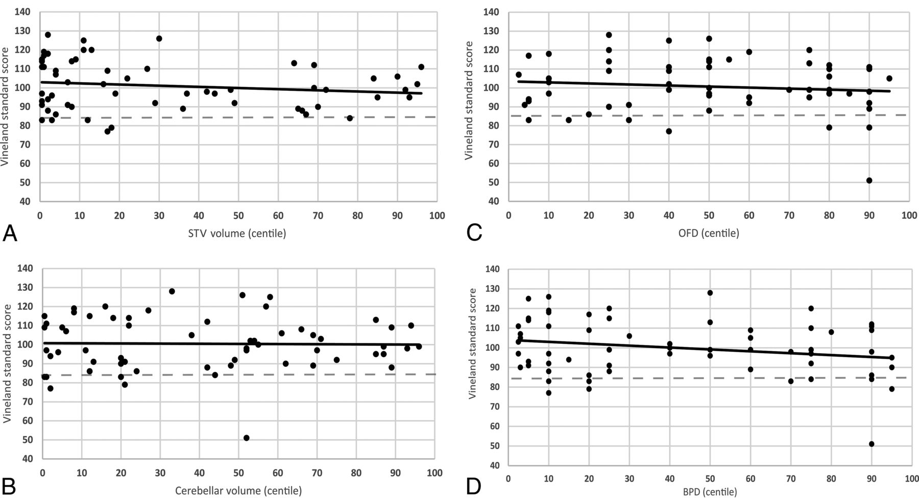
- FIG 1.
Scatterplots of the Vineland II Adaptive Behavior standard score according to STV (A), CV (B), OFD (C), and BPD (D). Macrocephalic and normocephalic biometry.
- FIG 2.
Scatterplots of the Vineland II Adaptive Behavior standard score according to STV (A), CV (B), OFD (C), and BPD (D). Microcephalic and normocephalic biometry.


Tables
Statistics Maternal age at birth (median) (IQR) (yr) 34 (29.8–36.0) Fetal sex Male (No.) (%) 34 (48.6%) Female (No.) (%) 36 (51.4%) Abnormal triple test findings (No.) (%) 3 (4.3%) Abnormal nuchal translucency findings (No.) (%) 3 (4.3%) Karyotype (No.) (%) Not performed 31 (44.3%) Normal findings 39 (55.7%) Abnormal findings 0 (0%) Chromosomal microarray (No.) (%) Not performed 48 (68.6%) Normal findings 21 (30%) Abnormal findings 1 (1.4%) Length of pregnancy (mean) (SD) (wk) 38.8 (1.3) Characteristics Median IQR OFD (mm) 98.5 93.0–102.3 OFD (centile) 57.5 28.8–90.0 BPD (mm) 77.0 72.8–80.0 BPD (centile) 40.0 10.0–75.0 TCD (mm) 45.0 41.9–48.0 TCD (centile) 50.0 40.0–75.0 STV (mm3) 220.294 192,195–248,017 STV (centile) 28.0 4.0–84.3 CV (mm3) 12.683 10,604–15,114 CV (centile) 48.5 12.8–77.5 CV/STV 0.058 0.052–0.065 CV/STV (centile) 58.5 32.0–88.5 - Table 3:
Comparison of Vineland-II Adaptive Behavior standard scores between fetuses with microcephaly and healthy fetuses according to 2D and 3D fetal brain MR imaging biometrya
Percentile of biometric value 4–96 ≤3 P Value MR imaging 3D STV (No. of fetuses) 43 16 Adaptive behavior composite score 99 (109–90) 104 (116–91) .44 MR imaging 3D CV (No. of fetuses) 53 9 Adaptive behavior composite score 99 (111–91) 97 (111–83) .51 MR imaging 2D OFD (No. of fetuses) 58 1 Adaptive behavior composite score 99 (111–71) 107 NA MR imaging 2D BPD (No. of fetuses) 61 6 Adaptive behavior composite score 98 (111–89) 104 (108–95) .68 Note:—NA indicates not applicable.
↵a Continuous variables are presented as median (IQR). P value refers to the Mann-Whitney U test.
- Table 4:
Comparison of Vineland-II Adaptive Behavior standard scores between fetuses with macrocephaly and healthy fetuses according to 2D and 3D fetal brain MR imaging biometrya
Percentile of biometric value 4–96 ≥97 P Value MR imaging 3D STV (No. of fetuses) 43 11 Adaptive behavior composite standard score 99 (109–90) 98 (109–84) .31 MR imaging 3D CV (No. of fetuses) 53 8 Adaptive behavior composite standard score 99 (111–91) 94 (106–84) .19 MR imaging 2D OFD (No. of fetuses) 58 11 Adaptive behavior composite standard score 99 (111–71) 90 (108–86) .13 MR imaging 2D BPD (No. of fetuses) 61 3 Adaptive behavior composite standard score 98 (111–89) 102 .93 ↵a Continuous variables are presented as median (IQR). P value refers to the Mann-Whitney U test.


Jump to section
Related Articles
Cited By...
- No citing articles found.