
Article Figures & Data
Figures
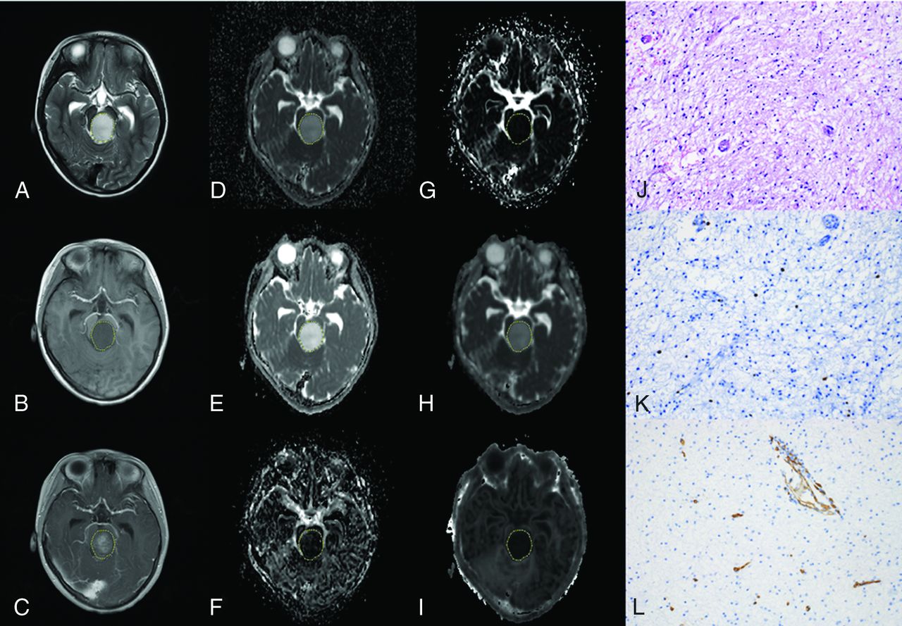
- FIG 1.
A 15-year-old boy with medulloblastoma in the cerebellum (WHO grade IV). The lesion shows hyperintensity on the T2-weighted image (A), hypointensity on the T1-weighted image (B), and enhancement on the postcontrast T1-weighted image (C). The lesion (VOI) demonstrates hypointensity on the ADC map (D), D map (E), and Dk map (H) and hyperintensity on the D* map (F), f map (G), and K map (I), with values of 0.647 × 10⁻³mm2/s, 0.594 × 10⁻³ mm2/s, 0.778 × 10⁻³ mm2/s, 87.228 × 10⁻³ mm2/s, 6.312%, and 1.210, respectively. The pathologic diagnosis was medulloblastoma with a cellularity of 4927 cell/mm2 (J), a Ki-67 index of 80% (K), and an MVD of 1.4% (L) (original magnification × 200).
- FIG 2.
A 5-year-old boy with a diffuse astrocytoma in the brain stem (WHO grade II). The lesion shows hyperintensity on the T2-weighted image (A), hypointensity on the T1-weighted image (B), and enhancement on the postcontrast T1-weighted image (C). The lesion (VOI) demonstrates hyperintensity on the ADC map (D), D map (E), and Dk map (H) and hypointensity on the D* map (F), f map (G), and K map (I), with values of 1.528× 10⁻³mm2/s, 1.530 × 10⁻³ mm2/s, 1.681 × 10⁻³ mm2/s, 57.310 × 10⁻³ mm2/s, 2.394%, and 0.315, respectively. The pathologic diagnosis was diffuse astrocytoma with a cellularity of 1917 cell/mm2 (J), a Ki-67 index of 1.1% (K), and an MVD of 0.9% (L) (magnification, × 200).
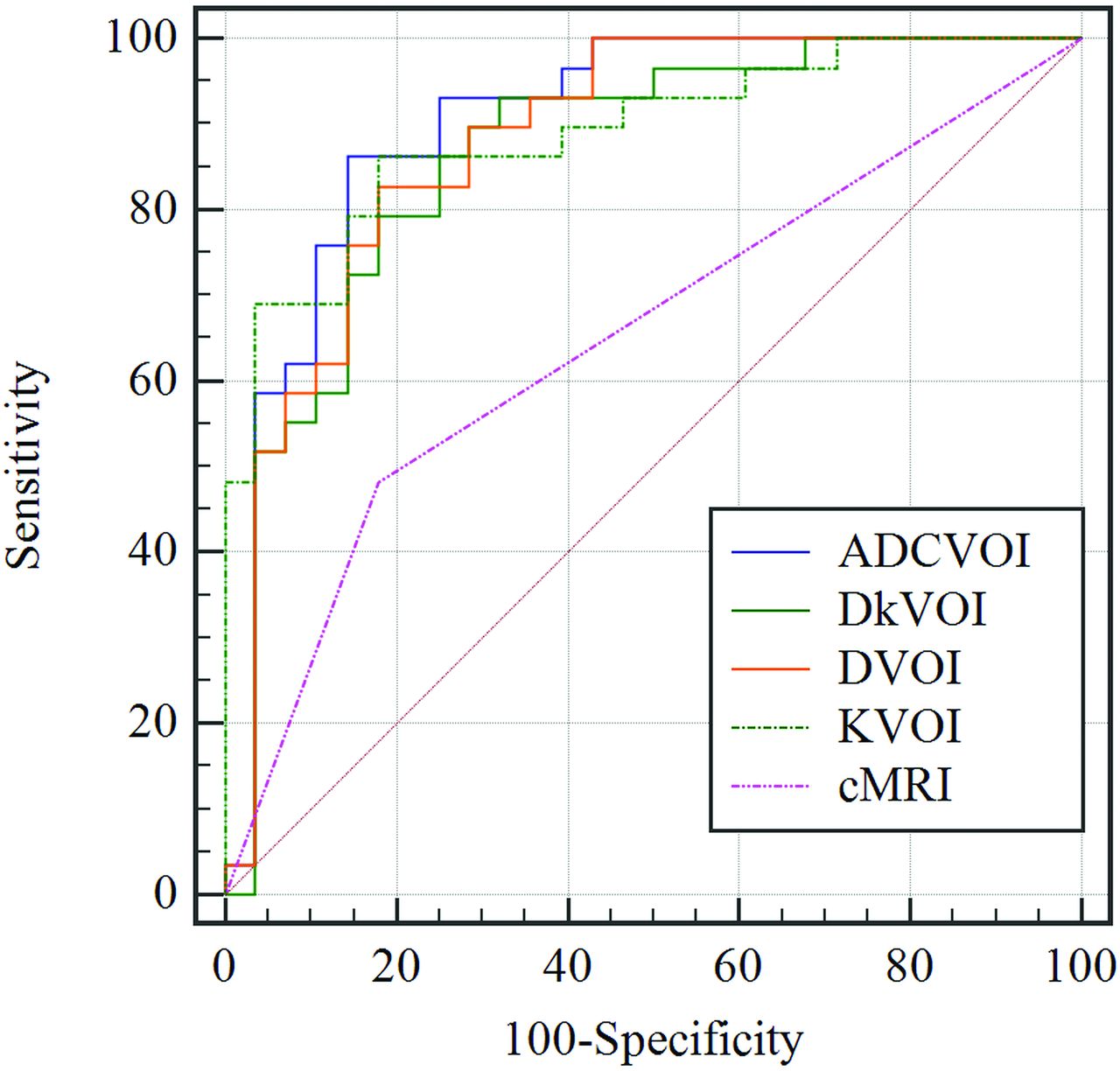
- FIG 3.
Receiver operating characteristic curves for ADCVOI, DVOI, DkVOI, KVOI, and conventional MR imaging in distinguishing low- from high-grade pediatric intracranial tumors.


Tables
- Table 1:
Comparison of demographic and conventional MR imaging characteristics between low- and high-grade PITs
Characteristics Low-Grade Tumor High-Grade Tumor P Value Demography Age (mean) (yr) 9.71 (SD, 3.66) 9.02 (SD, 4.11) .51 Male sex (No.) (%) 17 (65.4%) 23 (82.1%) .16 Location (No.) .13 Cerebral hemisphere 9 5 Cerebellum 12 11 Basal ganglia/brain stem 3 3 Other 2 9 Conventional MR imaging Cystic degeneration (No.) (%) 16 (61.5%) 13 (46.4%) .27 Hemorrhage (No.) (%) 5 (19.2%) 10 (35.7%) .18 Necrosis (No.) (%) 5 (19.2%) 14 (50.0%) .02 Enhancement (No.) (%) 21 (80.8%) 25 (89.3%) .62 Peritumoral edema (No.) (%) 10 (38.5%) 15 (53.6%) .27 Clear margin (No.) (%) 14 (53.8%) 13 (46.4%) .59 Parameters ICC (95% CI) Interreader Intrareader Conventional DWI parameters ADCROI (×10–3 mm2/s) 0.917 (0.862–0.951) 0.918 (0.863–0.951) ADCVOI (×10–3 mm2/s) 0.928 (0.879–0.957) 0.927 (0.878–0.957) IVIM parameters DROI (×1–3 mm2/s) 0.920 (0.867–0.953) 0.918 (0.862-0.951) DVOI (×10–3 mm2/s) 0.929 (0.880–0.958) 0.927 (0.876–0.957) D*ROI (×10–3 mm2/s) 0.771 (0.636–0.860) 0.807 (0.690–0.883) D*VOI (×10–3 mm2/s) 0.921 (0.869–0.954) 0.905 (0.842–0.944) fROI (%) 0.886 (0.812–0.932) 0.957 (0.926–0.975) fVOI (%) 0.934 (0.890–0.961) 0.898 (0.830–0.939) DKI parameters DkROI (×10-3 mm2/s) 0.928 (0.879-0.958) 0.926 (0.876-0.957) DkVOI (×10–3 mm2/s) 0.935 (0.891–0.962) 0.926 (0.876-0.957) KROI 0.982 (0.967–0.990) 0.984 (0.971–0.991) KVOI 0.983 (0.972–0.990) 0.984 (0.933–0.977) Note:—ICC indicates intraclass correlation coefficient.
- Table 3:
Comparison of histopathologic and quantitative MR imaging parameters between low- and high-grade PITsa
Parameters Low-Grade Tumor High-Grade Tumor P Value Conventional DWI parameters ADCROI (×10-3 mm2/s) 1.563 (1.275–1.732) 0.834 (0.735–1.269) <.001 ADCVOI (×10-3 mm2/s) 1.498 (1.254–1.692) 0.834 (0.725–1.118) <.001 IVIM parameters DROI (×10-3 mm2/s) 1.515 (1.233–1.724) 0.788 (0.660–1.246) <.001 DVOI (×10-3 mm2/s) 1.459 (1.224–1.677) 0.800 (0.668–1.081) <.001 D*ROI (×10-3 mm2/s) 82.962 (65.868–96.610) 87.105 (72.954–107.516) .36 D*VOI (×10-3 mm2/s) 81.562 (71.791–92.899) 81.271 (76.037–92.996) .43 fROI (%) (mean) 5.479 (SD, 2.603) 6.789 (SD, 2.773) .08 fVOI (%) (mean) 5.721 (SD, 2.183) 6.701 (SD, 2.852) .17 DKI parameters DkROI (×10-3 mm2/s) 1.899 (1.443–2.062) 1.053 (0.916–1.466) <.001 DkVOI (×10-3 mm2/s) 1.719 (1.440–2.004) 1.044 (0.835–1.353) <.001 KROI (mean) 0.483 (SD, 0.155) 0.887 (SD, 0.329) <.001 KVOI (mean) 0.500 (SD, 0.157) 0.912 (SD, 0.288) <.001 Histopathology (19 missing) Cellularity (mean) (cells/mm2) 2003 (SD, 769) 3175 (SD, 1161) .001 Ki-67 (%) 2.315 (0.945–5.310) 40.680 (21.195–66.310) <.001 MVD (%) 8.280 (4.340–15.400) 10.140 (8.345–19.995) .12 ↵a Data are expressed as mean (SD) or medians (lower quartile-upper quartile).
- Table 4:
Correlation between histologic parameters and quantitative MR imaging parameters for all PITs
Parameters Cellularity (r) (P Value) (Cells/mm2) Ki-67 (%) (r) (P Value) MVD (%) (r) (P Value) Conventional DWI parameters ADCROI (×10–3 mm2/s) –0.651 (P < .001) –0.717 (P < .001) 0.044 (P = .80) ADCVOI (×10–3 mm2/s) –0.659 (P < .001) –0.735 (P < .001) –0.031 (P = .86) IVIM parameters DROI (×10–3 mm2/s) –0.657 (P < .001) –0.714 (P < .001) 0.024 (P = .89) DVOI (×10–3 mm2/s) –0.657 (P < .001) –0.740 (P < .001) –0.021 (P = .91) D*ROI (×10–3 mm2/s) –0.161 (P = .36) –0.003 (P = .99) 0.082 (P = .64) D*VOI (×10–3 mm2/s) –0.029 (P = .87) 0.191 (P = .27) 0.273 (P = .11) fROI (%) 0.099 (P = .57) 0.140 (P = .42) 0.105 (P = .55) fVOI (%) 0.096 (P = .58) 0.269 (P = .12) 0.163 (P = .35) DKI parameters DkROI (×10–3 mm2/s) –0.548 (P < .001) –0.625 (P < .001) 0.122 (P = .49) DkVOI (×103 mm2/s) –0.601 (P < .001) –0.704 (P < .001) 0.061 (P = .73) KROI 0.677 (P < .001) 0.773 (P < .001) –0.101 (P = .56) KVOI 0.674 (P < .001) 0.802 (P < .001) –0.032 (P = .86) - Table 5:
Measurement of the quantitative MR imaging parameters and conventional MR imaging for differentiating high- and low-grade PITs
Parameters Cutoff Value Youden Index Sensitivity (%) Specificity (%) +LR (%) –LR (%) AUC ADCROI 1.238 0.558 75.0 80.8 3.90 0.31 0.826 ADCVOI 1.163 0.703 85.7 84.6 5.57 0.17 0.901 DROI 1.034 0.563 67.9 88.5 5.88 0.36 0.830 DVOI 1.119 0.668 82.1 84.9 5.34 0.21 0.894 DkROI 1.648 0.585 89.3 69.2 2.90 0.15 0.799 DKVOI 1.366 0.632 78.6 84.6 5.11 0.25 0.863 KROI 0.561 0.593 78.6 80.8 4.09 0.27 0.838 KVOI 0.593 0.665 85.7 80.8 4.46 0.18 0.885 cMRI 0.304 48.3 82.1 2.70 0.63 0.652 Note:—cMRI indicates conventional MRI; LR, likelihood ratio.


