Advertisement

Main menu

  • Home
  • Content
    • Current Issue
    • Accepted Manuscripts
    • Article Preview
    • Past Issue Archive
    • AJNR Case Collection
    • Case of the Week Archive
    • Classic Case Archive
    • Case of the Month Archive
  • Special Collections
    • Spinal CSF Leak Articles (Jan 2020-June 2024)
    • 2024 AJNR Journal Awards
    • Most Impactful AJNR Articles
  • Multimedia
    • AJNR Podcast
    • AJNR Scantastics
    • Video Articles
  • For Authors
    • Submit a Manuscript
    • Author Policies
    • Fast publishing of Accepted Manuscripts
    • Graphical Abstract Preparation
    • Manuscript Submission Guidelines
    • Imaging Protocol Submission
    • Submit a Case for the Case Collection
  • About Us
    • About AJNR
    • Editorial Board
  • More
    • Become a Reviewer/Academy of Reviewers
    • Subscribers
    • Permissions
    • Alerts
    • Feedback
    • Advertisers
    • ASNR Home
  • Other Publications
    • ajnr

User menu

  • Alerts
  • Log in

Search

  • Advanced search
American Journal of Neuroradiology

American Journal of Neuroradiology

ASHNR American Society of Functional Neuroradiology ASHNR American Society of Pediatric Neuroradiology ASSR
  • Alerts
  • Log in

Advanced Search

  • Home
  • Content
    • Current Issue
    • Accepted Manuscripts
    • Article Preview
    • Past Issue Archive
    • AJNR Case Collection
    • Case of the Week Archive
    • Classic Case Archive
    • Case of the Month Archive
  • Special Collections
    • Spinal CSF Leak Articles (Jan 2020-June 2024)
    • 2024 AJNR Journal Awards
    • Most Impactful AJNR Articles
  • Multimedia
    • AJNR Podcast
    • AJNR Scantastics
    • Video Articles
  • For Authors
    • Submit a Manuscript
    • Author Policies
    • Fast publishing of Accepted Manuscripts
    • Graphical Abstract Preparation
    • Manuscript Submission Guidelines
    • Imaging Protocol Submission
    • Submit a Case for the Case Collection
  • About Us
    • About AJNR
    • Editorial Board
  • More
    • Become a Reviewer/Academy of Reviewers
    • Subscribers
    • Permissions
    • Alerts
    • Feedback
    • Advertisers
    • ASNR Home
  • Follow AJNR on Twitter
  • Visit AJNR on Facebook
  • Follow AJNR on Instagram
  • Join AJNR on LinkedIn
  • RSS Feeds

Welcome to the new AJNR, Updated Hall of Fame, and more. Read the full announcements.


AJNR is seeking candidates for the position of Associate Section Editor, AJNR Case Collection. Read the full announcement.


Getting new auth cookie, if you see this message a lot, tell someone!
Research ArticleHead & Neck

The Cochlea in Branchio-Oto-Renal Syndrome: An Objective Method for the Diagnosis of Offset Cochlear Turns

A.F. Juliano, F. D’Arco, J. Pao, S. Picariello, E. Clement, G. Moonis and C.D. Robson
American Journal of Neuroradiology November 2022, 43 (11) 1646-1652; DOI: https://doi.org/10.3174/ajnr.A7653
A.F. Juliano
aFrom the Department of Radiology (A.F.J.), Massachusetts Eye and Ear, Harvard Medical School, Boston, Massachusetts
  • Find this author on Google Scholar
  • Find this author on PubMed
  • Search for this author on this site
  • ORCID record for A.F. Juliano
F. D’Arco
bDepartment of Radiology (F.D.), Great Ormond Street Hospital for Children, London, UK
  • Find this author on Google Scholar
  • Find this author on PubMed
  • Search for this author on this site
  • ORCID record for F. D’Arco
J. Pao
cDepartment of Radiology (J.P.), Orlando Health, Orlando Regional Medical Center, Orlando, Florida
  • Find this author on Google Scholar
  • Find this author on PubMed
  • Search for this author on this site
  • ORCID record for J. Pao
S. Picariello
dDepartment of General and Specialized Surgery for Women and Children (S.P.), University of Campania “Luigi Vanvitelli,” Naples, Italy
eNeuro-Oncology Unit (S.P.), Department of Paediatric Oncology, Santobono-Pausilipon Children’s Hospital, Naples, Italy
  • Find this author on Google Scholar
  • Find this author on PubMed
  • Search for this author on this site
  • ORCID record for S. Picariello
E. Clement
fDepartment of Clinical Genetics (E.C.), Great Ormond Street Hospital for Children National Health Service Foundation Trust, London, UK
  • Find this author on Google Scholar
  • Find this author on PubMed
  • Search for this author on this site
  • ORCID record for E. Clement
G. Moonis
gDepartment of Radiology (G.M.), NYU Langone Medical Center, New York, New York
  • Find this author on Google Scholar
  • Find this author on PubMed
  • Search for this author on this site
  • ORCID record for G. Moonis
C.D. Robson
hDepartment of Radiology (C.D.R.), Boston Children’s Hospital, Harvard Medical School, Boston, Massachusetts
  • Find this author on Google Scholar
  • Find this author on PubMed
  • Search for this author on this site
  • ORCID record for C.D. Robson
  • Article
  • Figures & Data
  • Info & Metrics
  • Responses
  • References
  • PDF
Loading

Article Figures & Data

Figures

  • Tables
  • FIG 1.
    • Download figure
    • Open in new tab
    • Download powerpoint
    FIG 1.

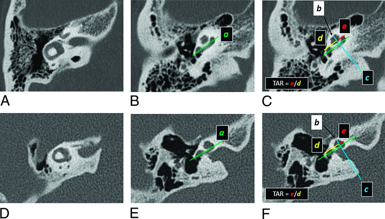
    Assessment of TAR of the cochlea on a patient without BOR (A–C) and a patient with EYA1-BOR (D–F). Standardized reformatted axial images are utilized, in which the planes are parallel to the plane of the lateral semicircular canal (A and D). Line a (green) is drawn parallel to the long axis of the basal turn (B and E), which is propagated across all axial images, including those where the apical or uppermost developed turn is visible (C and F). The midpoint of the uppermost turn is identified on the image that best displays it (black arrows in C and F); this point (point b) can also be propagated across all axial images. Line c (blue) is then drawn through point b (black arrow), perpendicular to line a (green). Distance d (between the anterior round window and the point of intersection, in yellow) and distance e (between medial bend of basal turn and the point of intersection, in red) are measured. TAR is e/d. As can be seen on these images, TAR in the patient with EYA1-BOR (F) is smaller than in the patient without BOR (C).

  • FIG 2.
    • Download figure
    • Open in new tab
    • Download powerpoint
    FIG 2.

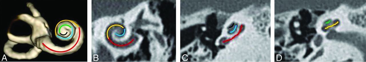
    A, Fifths of a cochlea, as demonstrated on a 3D reconstruction of the inner ear from a heavily T2-weighted sequence (3D DRIVE). The first fifth is in red (including the hook region), the second fifth in orange, the third fifth in blue, the fourth fifth in green, and the fifth fifth in black. (B, C, and D). CT of the temporal bone in a bone algorithm in the Stenvers view (B) and axial (C and D) planes shows the fifths of the cochlea in the same color scheme as depicted on the 3D model in A.

  • FIG 3.
    • Download figure
    • Open in new tab
    • Download powerpoint
    FIG 3.

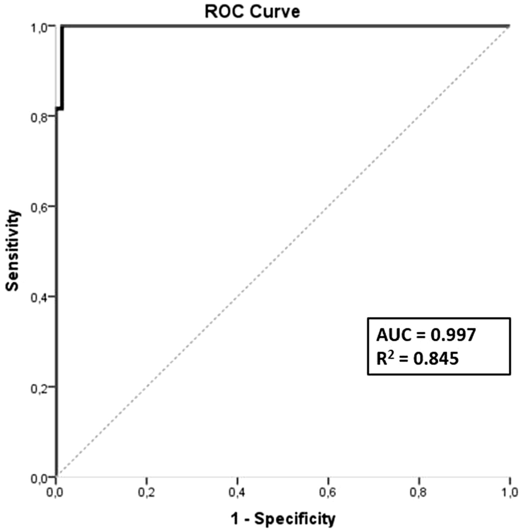
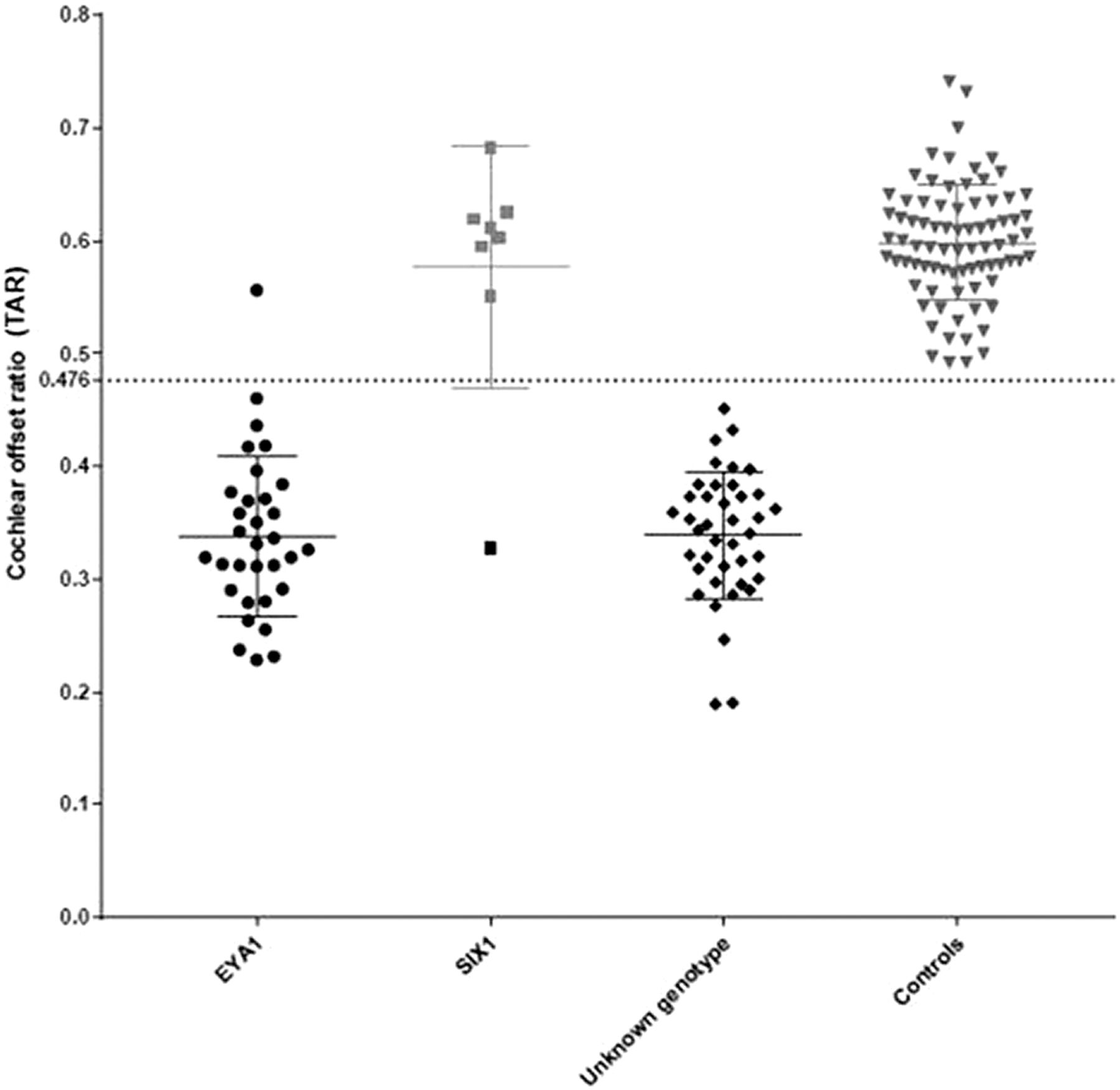
    Cochlear TAR among cochleae-deemed offset and not offset on visual assessment. EYA1-BOR: black round dots; SIX1-BOR: black square dots; unknown genotype: gray rhomboid dots; controls: gray triangle dots. The dashed line indicates the TAR cutoff (0.476) as determined by ROC curve analysis.

  • FIG 4.
    • Download figure
    • Open in new tab
    • Download powerpoint
    FIG 4.

    ROC curve.

  • FIG 5.
    • Download figure
    • Open in new tab
    • Download powerpoint
    FIG 5.

    Cochlear TAR among individuals with EYA1-BOR, SIX1-BOR, BOR of unknown genotype, and controls without BOR or sensorineural hearing loss. The TAR cutoff of 0.476 was determined by ROC curve analysis. All except one of the EYA1-BOR cochleae have TAR below the cutoff value. All except one of the SIX1-BOR cochleae have TAR above the cutoff value. All individuals with BOR of unknown genotype have TAR below the cutoff value. None of the controls have TAR below the cutoff value.

  • FIG 6.
    • Download figure
    • Open in new tab
    • Download powerpoint
    FIG 6.

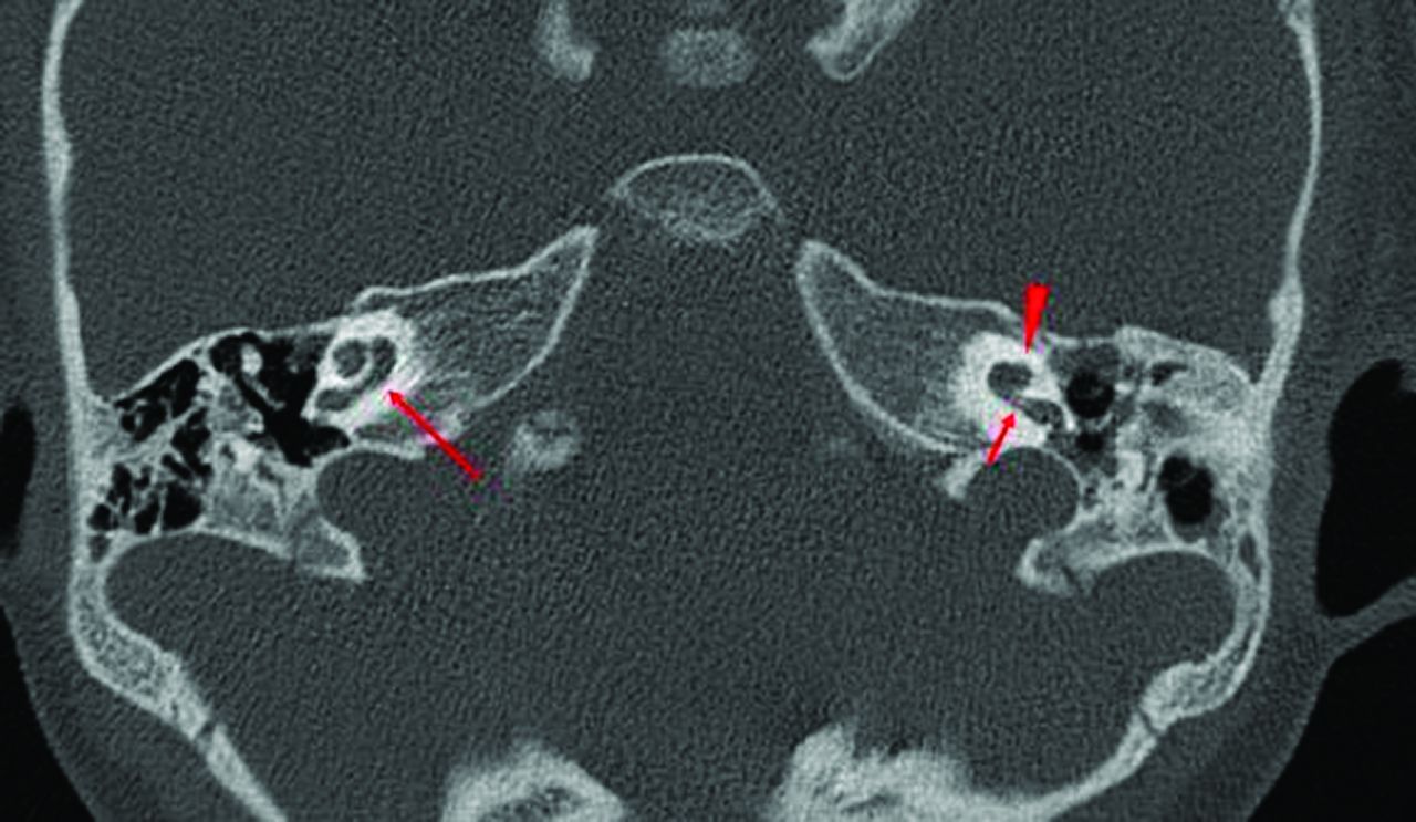
    Axial CT images of 3 different patients with EYA1-BOR showing the anteriorly offset unwound cochlea, an imaging feature characteristic of EYA1-BOR. Notice that the middle and apical turns are anteriorly located relative to the basal turn (red arrows) and slightly tilted away and separated from the basal turn (yellow arrowheads).

  • FIG 7.
    • Download figure
    • Open in new tab
    • Download powerpoint
    FIG 7.

    CT image of the patient with SIX1-BOR in whom the right cochlea does not demonstrate any offset (long arrow), while the left cochlea has an offset but with an appearance akin to cochlear hypoplasia type 4 rather than the typical unwound and offset cochlea of EYA1-BOR. Notice the normal size and morphology of the basal turn first half (short arrow), while the distal basal, middle, and apical turns are hypoplastic (arrowhead).

  • FIG 8.
    • Download figure
    • Open in new tab
    • Download powerpoint
    FIG 8.

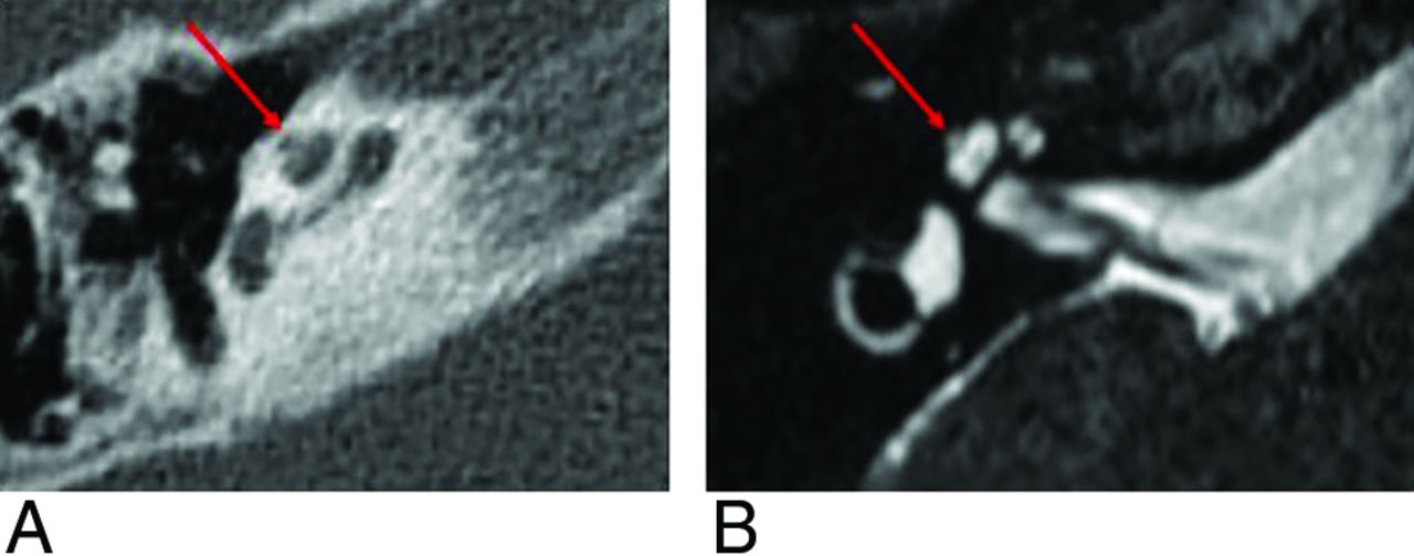
    A thorny apical turn in a patient with SIX1-BOR. The apical turn of the cochlea appears as a short, protuberant, thorny tip, as seen on CT (A) and MR imaging (B).

Tables

  • Figures
    • View popup
    Table 1:

    Qualitative (columns 3 and 4) and quantitative assessment (TAR, column 5) of cochlear offset

    Total No. of CochleaeNo. of Cochleae with OffsetNo. of Cochleae without OffsetMean TAR
    EYA1-BOR323200.338 (SD, 0.071)
    SIX1-BOR8170.577 (SD, 0.107)
    Unknown genotype BOR404000.339 (SD, 0.057)
    Controls800800.598 (SD, 0.051)
    • View popup
    Table 2:

    The presence/absence of thorny tips among the 3 BOR populations and in controls

    Total No. of CochleaeNo. of Cochleae with Thorny Tips
    EYA1-BOR320
    SIX1-BOR87
    Unknown genotype BOR400
    Controls800
PreviousNext
Back to top

In this issue

American Journal of Neuroradiology: 43 (11)
American Journal of Neuroradiology
Vol. 43, Issue 11
1 Nov 2022
  • Table of Contents
  • Index by author
  • Complete Issue (PDF)
Advertisement
Print
Download PDF
Email Article

Thank you for your interest in spreading the word on American Journal of Neuroradiology.

NOTE: We only request your email address so that the person you are recommending the page to knows that you wanted them to see it, and that it is not junk mail. We do not capture any email address.

Enter multiple addresses on separate lines or separate them with commas.
The Cochlea in Branchio-Oto-Renal Syndrome: An Objective Method for the Diagnosis of Offset Cochlear Turns
(Your Name) has sent you a message from American Journal of Neuroradiology
(Your Name) thought you would like to see the American Journal of Neuroradiology web site.
CAPTCHA
This question is for testing whether or not you are a human visitor and to prevent automated spam submissions.
Cite this article
A.F. Juliano, F. D’Arco, J. Pao, S. Picariello, E. Clement, G. Moonis, C.D. Robson
The Cochlea in Branchio-Oto-Renal Syndrome: An Objective Method for the Diagnosis of Offset Cochlear Turns
American Journal of Neuroradiology Nov 2022, 43 (11) 1646-1652; DOI: 10.3174/ajnr.A7653

Citation Manager Formats

  • BibTeX
  • Bookends
  • EasyBib
  • EndNote (tagged)
  • EndNote 8 (xml)
  • Medlars
  • Mendeley
  • Papers
  • RefWorks Tagged
  • Ref Manager
  • RIS
  • Zotero
0 Responses
Respond to this article
Share
Bookmark this article
Cochlea in Branchio-Oto-Renal Syndrome Diagnosis
A.F. Juliano, F. D’Arco, J. Pao, S. Picariello, E. Clement, G. Moonis, C.D. Robson
American Journal of Neuroradiology Nov 2022, 43 (11) 1646-1652; DOI: 10.3174/ajnr.A7653
del.icio.us logo Twitter logo Facebook logo Mendeley logo
  • Tweet Widget
  • Facebook Like
  • Google Plus One
Purchase

Jump to section

  • Article
    • Abstract
    • ABBREVIATIONS:
    • MATERIALS AND METHODS
    • RESULTS
    • DISCUSSION
    • CONCLUSIONS
    • Footnotes
    • References
  • Figures & Data
  • Info & Metrics
  • Responses
  • References
  • PDF

Related Articles

  • No related articles found.
  • PubMed
  • Google Scholar

Cited By...

  • No citing articles found.
  • Crossref
  • Google Scholar

This article has not yet been cited by articles in journals that are participating in Crossref Cited-by Linking.

More in this TOC Section

  • WHO Classification Update: Nasal&Skull Base Tumors
  • Peritumoral Signal in Vestibular Schwannomas
  • Chondrosarcoma vs Synovial Chondromatosis: Imaging
Show more HEAD & NECK

Similar Articles

Advertisement

Indexed Content

  • Current Issue
  • Accepted Manuscripts
  • Article Preview
  • Past Issues
  • Editorials
  • Editors Choice
  • Fellow Journal Club
  • Letters to the Editor

Cases

  • Case Collection
  • Archive - Case of the Week
  • Archive - Case of the Month
  • Archive - Classic Case

Special Collections

  • Special Collections

Resources

  • News and Updates
  • Turn around Times
  • Submit a Manuscript
  • Author Policies
  • Manuscript Submission Guidelines
  • Evidence-Based Medicine Level Guide
  • Publishing Checklists
  • Graphical Abstract Preparation
  • Imaging Protocol Submission
  • Submit a Case
  • Become a Reviewer/Academy of Reviewers
  • Get Peer Review Credit from Publons

Multimedia

  • AJNR Podcast
  • AJNR SCANtastic
  • Video Articles

About Us

  • About AJNR
  • Editorial Board
  • Not an AJNR Subscriber? Join Now
  • Alerts
  • Feedback
  • Advertise with us
  • Librarian Resources
  • Permissions
  • Terms and Conditions

American Society of Neuroradiology

  • Not an ASNR Member? Join Now

© 2025 by the American Society of Neuroradiology All rights, including for text and data mining, AI training, and similar technologies, are reserved.
Print ISSN: 0195-6108 Online ISSN: 1936-959X

Powered by HighWire