Advertisement

Main menu

  • Home
  • Content
    • Current Issue
    • Accepted Manuscripts
    • Article Preview
    • Past Issue Archive
    • AJNR Case Collection
    • Case of the Week Archive
    • Classic Case Archive
    • Case of the Month Archive
  • Special Collections
    • Spinal CSF Leak Articles (Jan 2020-June 2024)
    • 2024 AJNR Journal Awards
    • Most Impactful AJNR Articles
  • Multimedia
    • AJNR Podcast
    • AJNR Scantastics
    • Video Articles
  • For Authors
    • Submit a Manuscript
    • Author Policies
    • Fast publishing of Accepted Manuscripts
    • Graphical Abstract Preparation
    • Manuscript Submission Guidelines
    • Imaging Protocol Submission
    • Submit a Case for the Case Collection
  • About Us
    • About AJNR
    • Editorial Board
  • More
    • Become a Reviewer/Academy of Reviewers
    • Subscribers
    • Permissions
    • Alerts
    • Feedback
    • Advertisers
    • ASNR Home
  • Other Publications
    • ajnr

User menu

  • Alerts
  • Log in

Search

  • Advanced search
American Journal of Neuroradiology

American Journal of Neuroradiology

ASHNR American Society of Functional Neuroradiology ASHNR American Society of Pediatric Neuroradiology ASSR
  • Alerts
  • Log in

Advanced Search

  • Home
  • Content
    • Current Issue
    • Accepted Manuscripts
    • Article Preview
    • Past Issue Archive
    • AJNR Case Collection
    • Case of the Week Archive
    • Classic Case Archive
    • Case of the Month Archive
  • Special Collections
    • Spinal CSF Leak Articles (Jan 2020-June 2024)
    • 2024 AJNR Journal Awards
    • Most Impactful AJNR Articles
  • Multimedia
    • AJNR Podcast
    • AJNR Scantastics
    • Video Articles
  • For Authors
    • Submit a Manuscript
    • Author Policies
    • Fast publishing of Accepted Manuscripts
    • Graphical Abstract Preparation
    • Manuscript Submission Guidelines
    • Imaging Protocol Submission
    • Submit a Case for the Case Collection
  • About Us
    • About AJNR
    • Editorial Board
  • More
    • Become a Reviewer/Academy of Reviewers
    • Subscribers
    • Permissions
    • Alerts
    • Feedback
    • Advertisers
    • ASNR Home
  • Follow AJNR on Twitter
  • Visit AJNR on Facebook
  • Follow AJNR on Instagram
  • Join AJNR on LinkedIn
  • RSS Feeds

Welcome to the new AJNR, Updated Hall of Fame, and more. Read the full announcements.


AJNR is seeking candidates for the position of Associate Section Editor, AJNR Case Collection. Read the full announcement.


Research ArticleAdult Brain
Open Access

Generative Adversarial Network–Enhanced Ultra-Low-Dose [18F]-PI-2620 τ PET/MRI in Aging and Neurodegenerative Populations

K.T. Chen, R. Tesfay, M.E.I. Koran, J. Ouyang, S. Shams, C.B. Young, G. Davidzon, T. Liang, M. Khalighi, E. Mormino and G. Zaharchuk
American Journal of Neuroradiology September 2023, 44 (9) 1012-1019; DOI: https://doi.org/10.3174/ajnr.A7961
K.T. Chen
aFrom the Department of Biomedical Engineering (K.T.C.), National Taiwan University, Taipei, Taiwan
bDepartment of Radiology (K.T.C., M.E.I.K., J.O., S.S., G.D., T.L., M.K., G.Z.), Stanford University, Stanford, California
  • Find this author on Google Scholar
  • Find this author on PubMed
  • Search for this author on this site
  • ORCID record for K.T. Chen
R. Tesfay
cMeharry Medical College (R.T.), Nashville, Tennessee
  • Find this author on Google Scholar
  • Find this author on PubMed
  • Search for this author on this site
  • ORCID record for R. Tesfay
M.E.I. Koran
bDepartment of Radiology (K.T.C., M.E.I.K., J.O., S.S., G.D., T.L., M.K., G.Z.), Stanford University, Stanford, California
  • Find this author on Google Scholar
  • Find this author on PubMed
  • Search for this author on this site
  • ORCID record for M.E.I. Koran
J. Ouyang
bDepartment of Radiology (K.T.C., M.E.I.K., J.O., S.S., G.D., T.L., M.K., G.Z.), Stanford University, Stanford, California
  • Find this author on Google Scholar
  • Find this author on PubMed
  • Search for this author on this site
  • ORCID record for J. Ouyang
S. Shams
bDepartment of Radiology (K.T.C., M.E.I.K., J.O., S.S., G.D., T.L., M.K., G.Z.), Stanford University, Stanford, California
  • Find this author on Google Scholar
  • Find this author on PubMed
  • Search for this author on this site
C.B. Young
dDepartment of Neurology and Neurological Sciences (C.B.Y., E.M.), Stanford University, Stanford, California
  • Find this author on Google Scholar
  • Find this author on PubMed
  • Search for this author on this site
  • ORCID record for C.B. Young
G. Davidzon
bDepartment of Radiology (K.T.C., M.E.I.K., J.O., S.S., G.D., T.L., M.K., G.Z.), Stanford University, Stanford, California
  • Find this author on Google Scholar
  • Find this author on PubMed
  • Search for this author on this site
  • ORCID record for G. Davidzon
T. Liang
bDepartment of Radiology (K.T.C., M.E.I.K., J.O., S.S., G.D., T.L., M.K., G.Z.), Stanford University, Stanford, California
  • Find this author on Google Scholar
  • Find this author on PubMed
  • Search for this author on this site
M. Khalighi
bDepartment of Radiology (K.T.C., M.E.I.K., J.O., S.S., G.D., T.L., M.K., G.Z.), Stanford University, Stanford, California
  • Find this author on Google Scholar
  • Find this author on PubMed
  • Search for this author on this site
  • ORCID record for M. Khalighi
E. Mormino
dDepartment of Neurology and Neurological Sciences (C.B.Y., E.M.), Stanford University, Stanford, California
  • Find this author on Google Scholar
  • Find this author on PubMed
  • Search for this author on this site
  • ORCID record for E. Mormino
G. Zaharchuk
bDepartment of Radiology (K.T.C., M.E.I.K., J.O., S.S., G.D., T.L., M.K., G.Z.), Stanford University, Stanford, California
  • Find this author on Google Scholar
  • Find this author on PubMed
  • Search for this author on this site
  • ORCID record for G. Zaharchuk
  • Article
  • Figures & Data
  • Supplemental
  • Info & Metrics
  • Responses
  • References
  • PDF
Loading

Article Figures & Data

Figures

  • Tables
  • FIG 1.
    • Download figure
    • Open in new tab
    • Download powerpoint
    FIG 1.

    A schematic of the GAN (generator network, upper image; discriminator network, lower image) used in this work and its input and output channels. The arrows denote computational operations, and the tensors are denoted by boxes, with the number of channels indicated above each box. BN indicates batch normalization; Conv, convolution; Max, maximum; ReLU, rectified linear unit; tanh, hyperbolic tangent.
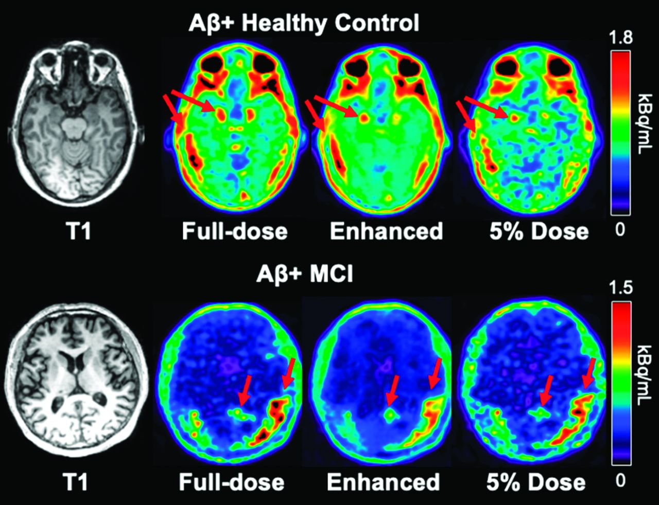
  • FIG 2.
    • Download figure
    • Open in new tab
    • Download powerpoint
    FIG 2.

    Representative τ PET images and their corresponding T1-weighted MR image in 2 individuals positive for amyloid. The enhanced PET image shows greatly reduced noise compared with the ultra-low-dose PET image. Arrows correspond to regions of abnormal elevated τ uptake. MCI indicates mild cognitive impairment.

  • FIG 3.
    • Download figure
    • Open in new tab
    • Download powerpoint
    FIG 3.

    Image-quality metrics comparing the ultra-low-dose PET (LD) and the ultra-low-dose enhanced PET (E) images with the ground truth full-dose PET image. PSNR indicates peak signal-to-noise ratio; SSIM, structural similarity; RMSE, root mean square error.
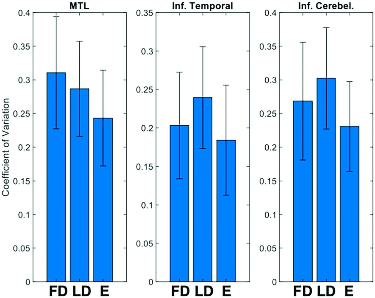
  • FIG 4.
    • Download figure
    • Open in new tab
    • Download powerpoint
    FIG 4.

    Mean (SD) of SUVR coefficient of variation in selected brain regions. E indicates enhanced images; FD, full-dose images; Inf. Cerebel, inferior cerebellum; MTL, medial temporal lobe; LD, ultra-low-dose image; Inf. Temporal, inferior temporal cortex
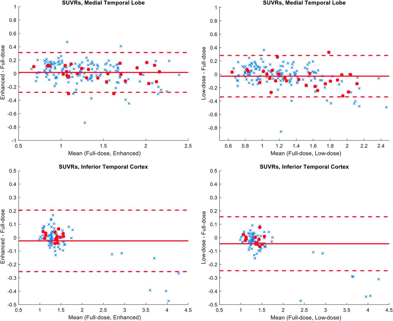
  • FIG 5.
    • Download figure
    • Open in new tab
    • Download powerpoint
    FIG 5.

    Bland-Altman plots comparing mean SUVRs in the ultra-low-dose PET and the enhanced PET with the full-dose PET images. The red dots denote healthy controls positive for amyloid, and the regions selected are the FreeSurfer labels, which make up the bilateral medial temporal lobe (entorhinal, amygdala) and the bilateral inferior temporal cortex.
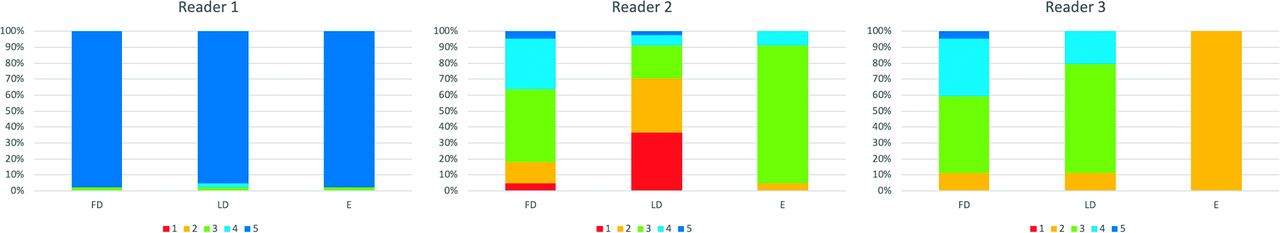
  • FIG 6.
    • Download figure
    • Open in new tab
    • Download powerpoint
    FIG 6.

    Quality scores of different image types as rated by 3 expert readers. Image quality scores: 1, uninterpretable; 2, bad; 3, adequate; 4, good; 5, excellent. FD indicates full-dose; LD, ultra-low-dose; E, enhanced.

Tables

  • Figures
    • View popup
    Table 1:

    Demographics and clinical indications of study population

    Healthy ControlADCBSMCIPSPsvPPA
    No.3151412
    Age (mean) (yr)70.13 (SD, 6.43)67.8 (SD, 13.48)7671.5 (SD, 10.54)7166, 78
    Sex (female)1230201
    Amyloid status7 P, 21 N4 P3 P, 1 N
    • Note:—CBS indicates cortical basal syndrome; MCI, mild cognitive impairment; N, negative; P, positive; PSP, progressive supranuclear palsy; svPPA, semantic variant primary-progressive aphasia.

    • View popup
    Table 2:

    Gwet's AC1 between and within readers of 10 randomly selected full-dose images on the tracer uptake in relevant brain regions and on the subjective image quality

    Gwet's AC1InterreaderReproducibility (Reader 1)Reproducibility (Reader 2)Reproducibility (Reader 3)
    Normal0.6770.7270.6281
    Entorhinal cortex0.813110.883
    Hipp./Amyg./Parahipp.0.6770.77111
    Inferior/mesial temporal0.901111
    Other cortex0.8300.8720.6691
    Primary eloquent cortex0.967111
    Quality (high vs low)0.79710.5990.760
    • Note:—Normal indicates no abnormal uptake in any of the selected regions; Hipp., hippocampus; Amyg., amygdala; Parahipp., parahippocampal gyrus.

    • View popup
    Table 3:

    Accuracy, sensitivity, and specificity of ultra-low-dose images and enhanced images compared with the full-dose images

    MetricFull-Dose vs Ultra-Low-DoseFull-Dose vs Enhanced
    Accuracy (%)Sensitivity (%)Specificity (%)Accuracy (%)Sensitivity (%)Specificity (%)
    Normal86.489.777.184.887.677.1
    Entorhinal cortex92.450.097.593.942.998.3
    Hipp./Amyg./Parahipp.87.954.295.488.666.793.5
    Inferior/mesial temporal95.583.397.493.277.895.6
    Other cortex87.973.990.888.687.089.0
    Primary eloquent cortex95.580.096.199.280.0100
    • Note:— Normal indicates no abnormal uptake in any of the selected regions; Hipp., hippocampus; Amyg., amygdala; Parahipp., parahippocampal gyrus.

PreviousNext
Back to top

In this issue

American Journal of Neuroradiology: 44 (9)
American Journal of Neuroradiology
Vol. 44, Issue 9
1 Sep 2023
  • Table of Contents
  • Index by author
  • Complete Issue (PDF)
Advertisement
Print
Download PDF
Email Article

Thank you for your interest in spreading the word on American Journal of Neuroradiology.

NOTE: We only request your email address so that the person you are recommending the page to knows that you wanted them to see it, and that it is not junk mail. We do not capture any email address.

Enter multiple addresses on separate lines or separate them with commas.
Generative Adversarial Network–Enhanced Ultra-Low-Dose [18F]-PI-2620 τ PET/MRI in Aging and Neurodegenerative Populations
(Your Name) has sent you a message from American Journal of Neuroradiology
(Your Name) thought you would like to see the American Journal of Neuroradiology web site.
CAPTCHA
This question is for testing whether or not you are a human visitor and to prevent automated spam submissions.
Cite this article
K.T. Chen, R. Tesfay, M.E.I. Koran, J. Ouyang, S. Shams, C.B. Young, G. Davidzon, T. Liang, M. Khalighi, E. Mormino, G. Zaharchuk
Generative Adversarial Network–Enhanced Ultra-Low-Dose [18F]-PI-2620 τ PET/MRI in Aging and Neurodegenerative Populations
American Journal of Neuroradiology Sep 2023, 44 (9) 1012-1019; DOI: 10.3174/ajnr.A7961

Citation Manager Formats

  • BibTeX
  • Bookends
  • EasyBib
  • EndNote (tagged)
  • EndNote 8 (xml)
  • Medlars
  • Mendeley
  • Papers
  • RefWorks Tagged
  • Ref Manager
  • RIS
  • Zotero
0 Responses
Respond to this article
Share
Bookmark this article
Ultra-Low-Dose [18F]-PI-2620 PET/MRI in Aging
K.T. Chen, R. Tesfay, M.E.I. Koran, J. Ouyang, S. Shams, C.B. Young, G. Davidzon, T. Liang, M. Khalighi, E. Mormino, G. Zaharchuk
American Journal of Neuroradiology Sep 2023, 44 (9) 1012-1019; DOI: 10.3174/ajnr.A7961
del.icio.us logo Twitter logo Facebook logo Mendeley logo
  • Tweet Widget
  • Facebook Like
  • Google Plus One
Purchase

Jump to section

  • Article
    • Abstract
    • ABBREVIATIONS:
    • MATERIALS AND METHODS
    • RESULTS
    • DISCUSSION
    • CONCLUSIONS
    • Footnotes
    • References
  • Figures & Data
  • Supplemental
  • Info & Metrics
  • Responses
  • References
  • PDF

Related Articles

  • PubMed
  • Google Scholar

Cited By...

  • A machine learning-based prediction of tau load and distribution in Alzheimers disease using plasma, MRI and clinical variables
  • Crossref (4)
  • Google Scholar

This article has been cited by the following articles in journals that are participating in Crossref Cited-by Linking.

  • Current Trends and Applications of PET/MRI Hybrid Imaging in Neurodegenerative Diseases and Normal Aging
    Jonathan Lee, Jonathan Renslo, Kasen Wong, Thomas G. Clifford, Bryce D. Beutler, Paul E. Kim, Ali Gholamrezanezhad
    Diagnostics 2024 14 6
  • A 3D multi-scale CycleGAN framework for generating synthetic PETs from MRIs for Alzheimer's disease diagnosis
    M. Khojaste-Sarakhsi, Seyedhamidreza Shahabi Haghighi, S.M.T. Fatemi Ghomi, Elena Marchiori
    Image and Vision Computing 2024 146
  • Machine learning prediction of tau‐PET in Alzheimer's disease using plasma, MRI, and clinical data
    Linda Karlsson, Jacob Vogel, Ida Arvidsson, Kalle Åström, Olof Strandberg, Jakob Seidlitz, Richard A. I. Bethlehem, Erik Stomrud, Rik Ossenkoppele, Nicholas J. Ashton, Henrik Zetterberg, Kaj Blennow, Sebastian Palmqvist, Ruben Smith, Shorena Janelidze, Renaud La Joie, Gil D. Rabinovici, Alexa Pichet Binette, Niklas Mattsson‐Carlgren, Oskar Hansson
    Alzheimer's & Dementia 2025 21 2
  • [18F]PI-2620 Tau PET signal across the aging and Alzheimer’s disease clinical spectrum
    Christina B. Young, Hillary Vossler, America Romero, Viktorija Smith, Jennifer Park, Alexandra N. Trelle, Joseph R. Winer, Edward N. Wilson, Michael M. Zeineh, Sharon J. Sha, Mehdi Khalighi, Maya V. Yutsis, Aimara P. Morales, David Anders, Greg Zaharchuk, Victor W. Henderson, Katrin I. Andreasson, Anthony D. Wagner, Kathleen L. Poston, Guido A. Davidzon, Elizabeth C. Mormino
    Imaging Neuroscience 2024 2

More in this TOC Section

  • Diagnostic Neuroradiology of Monoclonal Antibodies
  • Cerebral ADC Changes in Fabry Disease
  • ML for Glioma Molecular Subtype Prediction
Show more Adult Brain

Similar Articles

Advertisement

Indexed Content

  • Current Issue
  • Accepted Manuscripts
  • Article Preview
  • Past Issues
  • Editorials
  • Editors Choice
  • Fellow Journal Club
  • Letters to the Editor

Cases

  • Case Collection
  • Archive - Case of the Week
  • Archive - Case of the Month
  • Archive - Classic Case

Special Collections

  • Special Collections

Resources

  • News and Updates
  • Turn around Times
  • Submit a Manuscript
  • Author Policies
  • Manuscript Submission Guidelines
  • Evidence-Based Medicine Level Guide
  • Publishing Checklists
  • Graphical Abstract Preparation
  • Imaging Protocol Submission
  • Submit a Case
  • Become a Reviewer/Academy of Reviewers
  • Get Peer Review Credit from Publons

Multimedia

  • AJNR Podcast
  • AJNR SCANtastic
  • Video Articles

About Us

  • About AJNR
  • Editorial Board
  • Not an AJNR Subscriber? Join Now
  • Alerts
  • Feedback
  • Advertise with us
  • Librarian Resources
  • Permissions
  • Terms and Conditions

American Society of Neuroradiology

  • Not an ASNR Member? Join Now

© 2025 by the American Society of Neuroradiology All rights, including for text and data mining, AI training, and similar technologies, are reserved.
Print ISSN: 0195-6108 Online ISSN: 1936-959X

Powered by HighWire