Advertisement

Main menu

  • Home
  • Content
    • Current Issue
    • Accepted Manuscripts
    • Article Preview
    • Past Issue Archive
    • AJNR Case Collection
    • Case of the Week Archive
    • Classic Case Archive
    • Case of the Month Archive
  • Special Collections
    • Spinal CSF Leak Articles (Jan 2020-June 2024)
    • 2024 AJNR Journal Awards
    • Most Impactful AJNR Articles
  • Multimedia
    • AJNR Podcast
    • AJNR Scantastics
    • Video Articles
  • For Authors
    • Submit a Manuscript
    • Author Policies
    • Fast publishing of Accepted Manuscripts
    • Graphical Abstract Preparation
    • Manuscript Submission Guidelines
    • Imaging Protocol Submission
    • Submit a Case for the Case Collection
  • About Us
    • About AJNR
    • Editorial Board
  • More
    • Become a Reviewer/Academy of Reviewers
    • Subscribers
    • Permissions
    • Alerts
    • Feedback
    • Advertisers
    • ASNR Home
  • Other Publications
    • ajnr

User menu

  • Alerts
  • Log in

Search

  • Advanced search
American Journal of Neuroradiology

American Journal of Neuroradiology

ASHNR American Society of Functional Neuroradiology ASHNR American Society of Pediatric Neuroradiology ASSR
  • Alerts
  • Log in

Advanced Search

  • Home
  • Content
    • Current Issue
    • Accepted Manuscripts
    • Article Preview
    • Past Issue Archive
    • AJNR Case Collection
    • Case of the Week Archive
    • Classic Case Archive
    • Case of the Month Archive
  • Special Collections
    • Spinal CSF Leak Articles (Jan 2020-June 2024)
    • 2024 AJNR Journal Awards
    • Most Impactful AJNR Articles
  • Multimedia
    • AJNR Podcast
    • AJNR Scantastics
    • Video Articles
  • For Authors
    • Submit a Manuscript
    • Author Policies
    • Fast publishing of Accepted Manuscripts
    • Graphical Abstract Preparation
    • Manuscript Submission Guidelines
    • Imaging Protocol Submission
    • Submit a Case for the Case Collection
  • About Us
    • About AJNR
    • Editorial Board
  • More
    • Become a Reviewer/Academy of Reviewers
    • Subscribers
    • Permissions
    • Alerts
    • Feedback
    • Advertisers
    • ASNR Home
  • Follow AJNR on Twitter
  • Visit AJNR on Facebook
  • Follow AJNR on Instagram
  • Join AJNR on LinkedIn
  • RSS Feeds

Welcome to the new AJNR, Updated Hall of Fame, and more. Read the full announcements.


AJNR is seeking candidates for the position of Associate Section Editor, AJNR Case Collection. Read the full announcement.


Research ArticleBRAIN

Diffusion Tensor Imaging in Chronic Subdural Hematoma: Correlation between Clinical Signs and Fractional Anisotropy in the Pyramidal Tract

K. Yokoyama, M. Matsuki, H. Shimano, S. Sumioka, T. Ikenaga, K. Hanabusa, S. Yasuda, H. Inoue, T. Watanabe, M. Miyashita, R. Hiramatsu, K. Murao, A. Kondo, H. Tanabe and T. Kuroiwa
American Journal of Neuroradiology June 2008, 29 (6) 1159-1163; DOI: https://doi.org/10.3174/ajnr.A1001
K. Yokoyama
  • Find this author on Google Scholar
  • Find this author on PubMed
  • Search for this author on this site
M. Matsuki
  • Find this author on Google Scholar
  • Find this author on PubMed
  • Search for this author on this site
H. Shimano
  • Find this author on Google Scholar
  • Find this author on PubMed
  • Search for this author on this site
S. Sumioka
  • Find this author on Google Scholar
  • Find this author on PubMed
  • Search for this author on this site
T. Ikenaga
  • Find this author on Google Scholar
  • Find this author on PubMed
  • Search for this author on this site
K. Hanabusa
  • Find this author on Google Scholar
  • Find this author on PubMed
  • Search for this author on this site
S. Yasuda
  • Find this author on Google Scholar
  • Find this author on PubMed
  • Search for this author on this site
H. Inoue
  • Find this author on Google Scholar
  • Find this author on PubMed
  • Search for this author on this site
T. Watanabe
  • Find this author on Google Scholar
  • Find this author on PubMed
  • Search for this author on this site
M. Miyashita
  • Find this author on Google Scholar
  • Find this author on PubMed
  • Search for this author on this site
R. Hiramatsu
  • Find this author on Google Scholar
  • Find this author on PubMed
  • Search for this author on this site
K. Murao
  • Find this author on Google Scholar
  • Find this author on PubMed
  • Search for this author on this site
A. Kondo
  • Find this author on Google Scholar
  • Find this author on PubMed
  • Search for this author on this site
H. Tanabe
  • Find this author on Google Scholar
  • Find this author on PubMed
  • Search for this author on this site
T. Kuroiwa
  • Find this author on Google Scholar
  • Find this author on PubMed
  • Search for this author on this site
  • Article
  • Figures & Data
  • Info & Metrics
  • Responses
  • References
  • PDF
Loading

Abstract

BACKGROUND AND PURPOSE: Diffusion tensor imaging (DTI) was introduced as a good technique to evaluate structural abnormalities in the white matter. In this study, we used DTI to examine anisotropic changes of the pyramidal tracts displaced by chronic subdural hematoma (CSDH).

MATERIALS AND METHODS: Twenty-six patients with unilateral CSDH underwent DTI before and after surgery. We measured fractional anisotropy (FA) values in pyramidal tracts of bilateral cerebral peduncles and calculated the ratio of the FA value on the lesion side to that on the contralateral side (FA ratio) and compared the ratios with motor weakness. Moreover, the relationships between FA ratios and clinical factors such as age, sex, midline shift, interval from trauma, and hematoma attenuation on CT were evaluated.

RESULTS: FA values of pyramidal tracts on the lesion side were significantly lower than those on the contralateral side (0.66 ± 0.07 versus 0.74 ± 0.05, P < .0001). The FA ratio was correlated to the severity of motor weakness (r2 = 0.32, P = .002). FA ratios after surgery improved significantly compared with those before surgery (0.96 ± 0.08 versus 0.89 ± 0.07, P = .0004). Intervals from trauma and the midline shift were significantly associated with decreased FA ratios (P = .0008 and P = .037).

CONCLUSIONS: In patients with CSDH, a reversible decrease of FA in the affected pyramidal tract on DTI was correlated to motor weakness. These anisotropic changes were considered to be caused by a reversible distortion of neuron fibers and vasogenic edema due to the hematoma.

Chronic subdural hematoma (CSDH) is a progressive space-occupying lesion, which may lead to reversible reduction in cerebral function due to brain compression or brain displacement. The causes, clinical characteristics, and therapeutic management of CSDH are well established; however, the detailed physiology, including the mechanisms of appearance with motor dysfunction in patients, is still controversial.1–4

Diffusion tensor imaging (DTI) reveals the tissue microstructure and architecture of white matter tracts, including pyramidal tracts, and extracts the principal diffusivities, mean diffusivities (MD), and fractional anisotropy (FA). Previously, it was reported that decreased FA in the pyramidal tracts on DTI was associated with secondary degeneration and could be a reliable measure of motor weakness.5–8 The purpose of this study was to investigate changes of DTI in affected pyramidal tracts of patients with CSDH and to evaluate the association between changes of FA and clinical factors such as age, sex, hematoma thickness, midline shift, interval from the trauma, and attenuation on CT.

Materials and Methods

Study Populations

From September 2006 to February 2007, 26 consecutive patients (17 men and 9 women; age range, 44–90 years; mean, 78 ± 7.8 years; 15 right side and 11 left side; maximal width of hematoma, 21.4 ± 4.1 mm) with unilateral CSDH were examined by MR imaging 1–2 days before and 6–8 days after hematoma removal by burr-hole craniotomy (Table 1). In this study, we excluded patients with bilateral CSDH and patients who had pre-existing neurologic diseases such as cerebral infarction or hematoma. Seventeen of 26 patients had motor weakness, with the Manual Muscle Test (MMT)9 scores as follows: grade 2 (the patient has all or partial range of motion in the gravity-eliminated position) in 1 patient, grade 3 (the patient can tolerate no resistance but can perform the movement through the full range of motion) in 3 patients, and grade 4 (the patient can hold the position against strong-to-moderate resistance and has full range of motion) in 13 patients.

View this table:
  • View inline
  • View popup
Table 1:

Patient details and clinical information

Immediately after burr-hole craniotomy, motor dysfunctions of 13 of 17 patients were drastically improved. The other 4 patients needed rehabilitation to be discharged from the hospital because of low-grade gait disturbance due to residual motor weakness (cases 5, 10, 18, and 21); these patients recovered within 2 weeks.

In comparison, 20 healthy volunteers (10 men and 10 women; age range, 13–74 years; mean, 54 ± 20 years) were examined by MR imaging in the same period. The study protocol was approved by the institutional review board, and informed consent was obtained from all patients.

MR Protocol

MR imaging was performed on a 3T MR imaging scanner (Signa Excite; GE Healthcare, Milwaukee, Wis) by using an 8-element phased-array neurovascular array coil. DTI was performed by using a single-shot echo-planar sequence with the array sensitivity–encoding technique. Motion-probing gradient orientations were applied along 15 directions, and the b factor was set at 1000 s/mm2. The acquisition parameters used were as follows: TR/TE, 12,000/70–76 ms; bandwidth, 143 kHz; matrix size, 128 × 128; section thickness, 3 mm without intersection gap; FOV, 30 × 30 cm; and NEX, 1. In addition, a reference image without diffusion weighting (b = 0 s/mm2) was obtained. In all patients and healthy volunteers, 50 sections were acquired parallel to the line of the nasofrontal angle and the pituitary fossa.

Image Analysis

One experienced neuroradiologist (M.M.), who was unaware of the neurologic status of the subjects, placed regions of interest 3 times in the anterior parts of the bilateral cerebral peduncle, which correspond to a pyramidal tract based on anatomic knowledge, of healthy volunteers and patients before and after surgery and measured and averaged the FA and MD values. Another experienced neurosurgeon (K.Y.) investigated the differences in FA and MD values between the right and left sides in healthy volunteers and between the lesion and the contralateral sides in patients before surgery (Fig 1).

Fig 1.
  • Download figure
  • Open in new tab
  • Download powerpoint
Fig 1.

A and B, Head CT and T2-weighted MR images of CSDH, respectively. C and D, Regions of interest in the bilateral pyramidal tract of the cerebral peduncle. C, FA map. D, Apparent diffusion coefficient map.

We calculated FA ratios (ratios of FA values in the lesion side/normal side) in the patients before surgery and investigated the correlation between the preoperative FA ratio and the presented motor weakness, which was classified by MMT. Furthermore, we analyzed the association between the preoperative FA ratio and clinical factors such as age, sex, hematoma thickness, midline shift, interval from the trauma, and attenuation on CT. Then we compared patients' FA ratios before and after surgery.

Statistical analyses were performed with the Wilcoxon signed rank test and a linear regression analysis by using StatView 5.0 software (SAS Institute, Cary, NC).10 All data are presented as means ± SD, and differences were considered statistically significant at a P value < .05.

Results

Patients' characteristics of this series are summarized in Table 1.

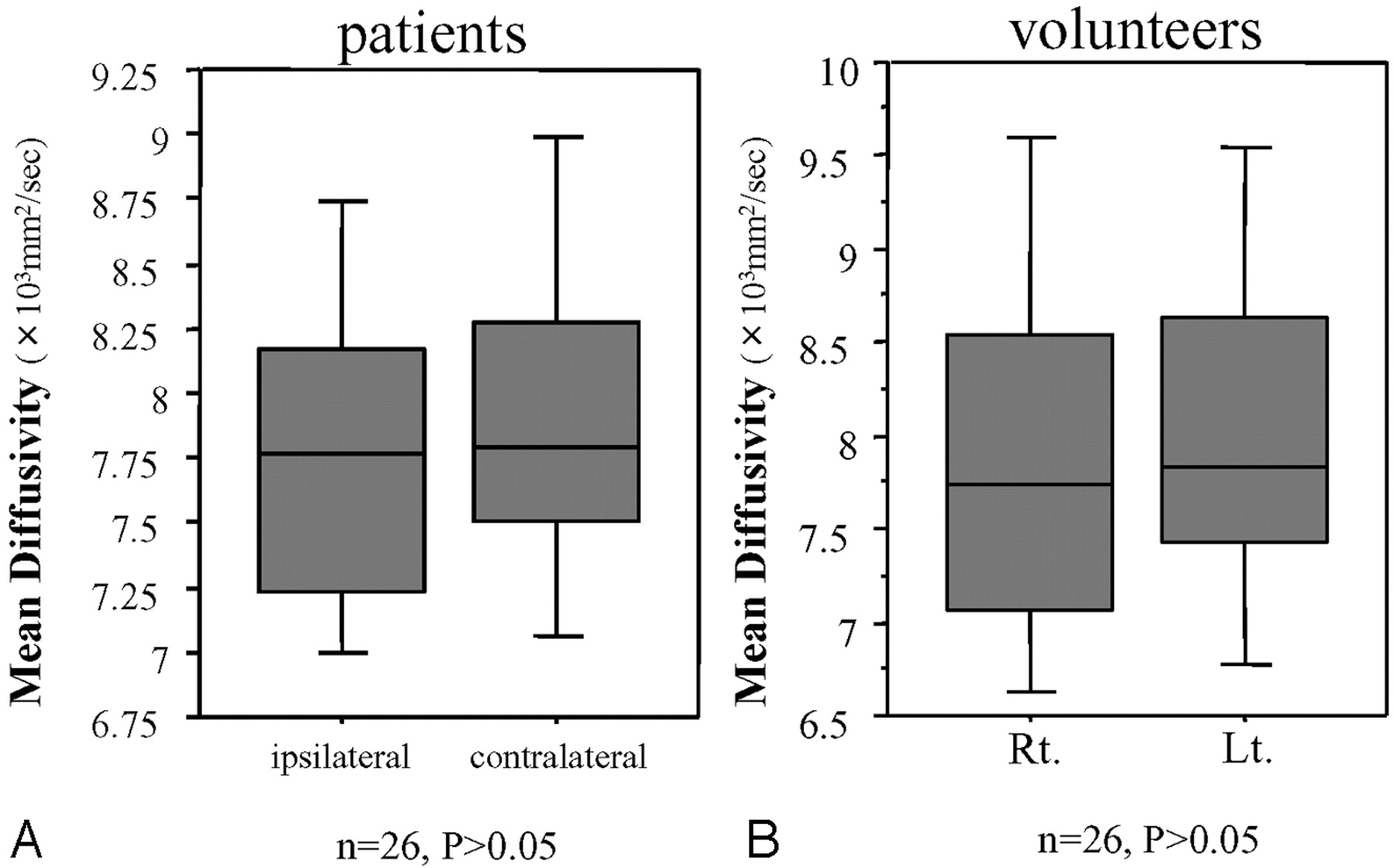
In healthy volunteers, there was no significant difference in FA values between bilateral pyramidal tracts (right versus left, 0.72 ± 0.05 versus 0.73 ± 0.03; P = .46). On the other hand, in patients with CSDH, the FA value of the pyramidal tract on the lesion side was significantly lower than that on the contralateral side (0.66 ± 0.07 versus 0.74 ± 0.05, P < .0001) (Fig. 2). With regard to the MD value, in patients and healthy volunteers, there was no significant difference in bilateral tracts (patients: 8.0 ± 0.8 10−4 mm2/s versus 8.1 ± 1.1 10−4 mm2/s, P > .05; volunteers: 7.9 ± 1.2 10−4 mm2/s versus 8.0 ± 1.1 10−4 mm2/s, P > .05) (Fig 3).

Fig 2.
  • Download figure
  • Open in new tab
  • Download powerpoint
Fig 2.

Region-of-interest analysis of FA values in the cerebral peduncle for lesion sides and contralateral sides. The Wilcoxon signed rank test was used for analysis of the differences. A, In patients with CSDH, FA values of the affected sides are significantly lower than those of intact sides (P < .0001). B, In healthy volunteers, there is no significant difference between bilateral sides (P > .5). Lt. indicates left; Rt, right.

Fig 3.
  • Download figure
  • Open in new tab
  • Download powerpoint
Fig 3.

Region-of-interest analysis of MD values in the cerebral peduncle for lesion sides and contralateral sides. The Wilcoxon signed rank test was used for analysis of the differences. A, In patients with CSDH, there is no significant difference in MD values between the affected sides and the contralateral sides (P > .5). B, In healthy volunteers, there is no significant difference between bilateral sides (P > .5). Lt. indicates left; Rt, right.

Linear regression analysis revealed that the preoperative FA ratio was significantly correlated to MMT (r2 = 0.32, P = .002) (Fig 4). The preoperative FA ratio was associated with the interval from the trauma and the midline shift determined on CT (interval from trauma, P = .0008; midline shift, P = .037). Age, hematoma thickness, and attenuation of the hematoma on CT were not associated with the FA ratio (P > .05) (Table 2). In 20 (87.0%) of 23 patients, the FA of the pyramidal tract on the lesion side after surgery increased significantly compared with that before the surgery (0.89 ± 0.08 versus 0.96 ± 0.08, P = .0004) (Fig 5). The other 3 patients (cases 14, 16, and 17) had no motor weakness before surgery.

Fig 4.
  • Download figure
  • Open in new tab
  • Download powerpoint
Fig 4.

The correlation between the initial FA ratio and motor weakness. Initial FA ratios are significantly correlated with motor weakness (R2 = 0.32, P = .002) by linear regression analysis. Preope. indicates preoperative.

Fig 5.
  • Download figure
  • Open in new tab
  • Download powerpoint
Fig 5.

The comparison FA ratios before and after burr-hole craniotomy in 23 patients with CSDH. FA ratios are significantly increased after surgery (Wilcoxon signed rank test, P = .0004). Pre-ope indicates preoperative; Post-ope, postoperative.

View this table:
  • View inline
  • View popup
Table 2:

Relationship between clinical data and FA ratio

Discussion

CSDH generally occurs in the elderly, with a reported annual incidence of 5.3 patients treated per 10,0000 individuals.11 In 60%–80% of patients, a traumatic event is reported to precede the hemorrhage,2,12 but a mild traumatic event may go unrecognized. Traumatic subdural effusion is a result of arachnoid tearing caused by head injury or neurosurgery, and this fluid with or without blood in the subdural space facilitates the formation of a medial membrane. This medial membrane then forms internal capillaries or sinusoids. These fenestrated blood vessels allow plasma fluid leakage into and resultant enlargement of the subdural space. Bleeding then occurs repeatedly from capillaries, with the degeneration of the endothelium, and is accompanied by local hyperfibrinolysis, which is one of the causes of the growth of effusion into the CSDH. Among our patients, the common symptoms were gait disturbance, hemiparesis, headache, and dementia. Generally, in cases with symptomatic or expanding hematoma, a burr-hole craniotomy is performed. Following the surgical treatment, most patients with CSDH immediately and dramatically recover their premorbid function. Some previous reports have demonstrated that a reversible reduction of cerebral function can be caused by a decreased cerebral blood flow or vasogenic edema in the affected pyramidal tract; however, the detailed physiology has remained controversial.13–15

In patients with CSDH, our results showed significantly reduced FA in the affected descending pyramidal tract on DTI, correlated with the severity of the motor dysfunction. These findings coincide with those of secondary degeneration caused by stroke, tumor, trauma, and so forth, as demonstrated in previous reports.16–21 Moreover, our results showed significant improvement of the FA ratio with a rapid relief of motor weakness after hematoma removal. Recently, some reports have demonstrated cases with reversible loss of anisotropy in the white matter. Kim et al8 and Yamada et al22 reported that a decreased FA value on DTI did not necessarily represent direct damage to the tract but was more commonly noted to be a result of vasogenic edema and tract compression by the mass effect. Then, they believed that reversible loss of FA in cases of CSDH is incurred by the distortion of the pyramidal tract and vasogenic edema due to expansion of the hematoma.

In the factor analysis, the initial FA ratio was significantly associated with the interval from trauma and the midline shift (Table 2). Although there was no statistical significance, FA ratios tended to decrease in patients with mixed-attenuation hematoma on CT. These results indicated that a rapidly growing hematoma will invite severe distortion and vasogenic edema, which result in a decrease of the FA ratio. Finally, motor deficits are accompanied by decreased FA in the pyramidal tract. However, the thickness of the hematoma was not associated with the decreased FA ratio. It was believed that a thick hematoma sometimes causes only a tiny distortion and vasogenic edema in elderly patients with atrophic cerebral hemispheres.

There are certain limitations to this study. First, the patient population was small, and the power of statistical analysis was limited. In the present study, patients with bilateral CSDH were excluded because of the need to compare FA values of pyramidal tracts between affected and nonaffected sides. Second, all demonstrated FA values were analyzed in the cerebral peduncles, and data of FA values in other parts of the pyramidal tracts were not shown. In the present study, regions of interest were defined on the basis of an anatomic knowledge of the location of the pyramidal tract. Beforehand, we analyzed FA values not only in the cerebral peduncles but also in the corona radiata and internal capsule. The reproducibility of the obtained FA values in the cerebral peduncles was the most consistent among the 3 areas, so we chose the cerebral peduncles for analysis in this study. In the future, DTI will play an important role in clarifying the detailed physiology of CSDH. A much larger population of patients with CSDH must be subjected to DTI to accomplish this clarification.

Conclusion

Using DTI, we confirmed a reversibility of the declined FA in the distal pyramidal tract of patients with CSDH. The reversible loss of FA was considered to be incurred by the distortion of the pyramidal tract and vasogenic edema due to expansion of the hematoma.

References

  1. ↵
    Markwalder TM, Steinsiepe KF, Rohner M, et al. The course of chronic subdural hematomas after burr-hole craniostomy and closed-system drainage. J Neurosurg 1981;55:390–96
  2. ↵
    Robinson RG: Chronic subdural hematoma: surgical treatment in 133 patients. J Neurosurg 1984;61:263–68
    PubMed
  3. Stroobandt G, Fransen P, Thauvoy C, et al. Pathogenic factors in chronic subdural hematoma and causes of recurrence after drainage. Acta Neurochir (Wien) 1995;137:6–14
    CrossRefPubMed
  4. ↵
    Victorotos GC, Bligh AS. A more systematic management of subdural hematoma with the aid of CT scan. Surg Neurol 1981;15:158–60
    CrossRefPubMed
  5. ↵
    Berman JI, Berger MS, Mukherjee P, et al. Diffusion-tensor imaging-guided tracking of fibers of the pyramidal tract combined with intraoperative cortical simulation mapping in patients with gliomas. J Neurosurg 2004;101:66–72
    PubMed
  6. Clark CA, Barrick TR, Murphy MM, et al. White matter fiber tracking in patients with space-occupying lesions of the brain: a new technique for neurosurgical planning? Neuroimage 2003;20:1602–08
  7. Hendler T, Pianka P, Sigal M, et al. Delineating gray and white matter involvement in brain lesions: three-dimensional alignment of functional magnetic resonance and diffusion tensor imaging. J Neurosurg 2003;99:1018–27
    PubMed
  8. ↵
    Kim CH, Chung CK, Kim JC, et al. Use of diffusion tensor imaging to evaluate weakness. J Neurosurg 2007;106:111–18
    CrossRefPubMed
  9. ↵
    Gonnella C. The manual muscle test in the patient's evaluation and program for treatment. Phys Ther Rev 1954;34:16–18
    PubMed
  10. ↵
    Altman DG. Practical Statistics for Medical Research. London: Chapman and Hall;1994 :403–09
  11. ↵
    Matsumoto M, Sakata Y, Yamazaki T, et al. Local coagulofibrinolysis in the postsurgical recovery of patients with chronic subdural hematoma. Acta Neurochir (Wien) 1999;141:177–81
    CrossRefPubMed
  12. ↵
    Nomura S, Kashiwagi S, Fujisawa H, et al. Characterization of local hyperfibrinolysis in chronic subdural hematomas by SDS-PAGE and immunoblot. J Neurosurg 1994;81:910–13
    PubMed
  13. ↵
    Yamada M, Suzuki A, Yasui N, et al. Analysis using positron emission topography (PET) in hemiparetic patients with chronic subdural hematoma. No Shinkei Geka 2002 :30:1053–57
    PubMed
  14. Inao S, Kawai T, Kabeya R, et al. Relation between brain displacement and local cerebral blood flow in patients with chronic subdural haematoma. J Neurol Neurosurg Psychiatry 2001;71:741–46
    Abstract/FREE Full Text
  15. ↵
    Kuwabara H. Regional cerebral blood flow and metabolism in chronic subdural hematoma. Neurosurg Clin N Am 2000;11:499–502
    PubMed
  16. ↵
    Liang Z, Zeng J, Liu S, et al. A prospective study of secondary degeneration following subcortical infarction using diffusion tensor imaging. J Neurol Neurosurg Psychiatry 2007;78:581–86. Epub 2007 Jan 19
    Abstract/FREE Full Text
  17. Werring DJ, Toosy AT, Clark CA, et al. Diffusion tensor imaging can detect and quantify corticospinal tract degeneration after stoke. J Neurol Neurosurg Psychiatry 2000;69:269–72
    Abstract/FREE Full Text
  18. Thomalla G, Glauche V, Koch MA, et al. Diffusion tensor imaging detects early wallerian degeneration of the pyramidal tract after ischemic stroke. Neuroimage 2004;22:1767–74
    CrossRefPubMed
  19. Kirton A, Shroff M, Visvanathan T, et al. Quantified corticospinal tract diffusion restriction predicts neonatal stroke outcome. Stroke 2007;38:974–80
    Abstract/FREE Full Text
  20. Moller M, Frandsen J, Anderson G, et al. Dynamic changes of corticospinal tracts after stroke detected by fiber tracking. J Neurol Neurosurg Psychiatry 2007;78:587 -92. Epub Jan 8
    Abstract/FREE Full Text
  21. ↵
    Huisman TA, Schwamm LH, Schaefer PW, et al. Diffusion tensor imaging as potential biomarker of white matter injury in diffuse axonal injury. AJNR Am J Neuroradiol 2004;25:370–76
    Abstract/FREE Full Text
  22. ↵
    Yamada K, Kizu O, Mori S, et al. Brain fiber tracking with clinically feasible diffusion-tensor MR imaging: initial experience. Radiology 2003;227:295–301
    CrossRefPubMed
  • Received October 23, 2007.
  • Accepted after revision December 23, 2007.
  • Copyright © American Society of Neuroradiology
View Abstract
PreviousNext
Back to top

In this issue

American Journal of Neuroradiology: 29 (6)
American Journal of Neuroradiology
Vol. 29, Issue 6
June 2008
  • Table of Contents
  • Index by author
Advertisement
Print
Download PDF
Email Article

Thank you for your interest in spreading the word on American Journal of Neuroradiology.

NOTE: We only request your email address so that the person you are recommending the page to knows that you wanted them to see it, and that it is not junk mail. We do not capture any email address.

Enter multiple addresses on separate lines or separate them with commas.
Diffusion Tensor Imaging in Chronic Subdural Hematoma: Correlation between Clinical Signs and Fractional Anisotropy in the Pyramidal Tract
(Your Name) has sent you a message from American Journal of Neuroradiology
(Your Name) thought you would like to see the American Journal of Neuroradiology web site.
CAPTCHA
This question is for testing whether or not you are a human visitor and to prevent automated spam submissions.
Cite this article
K. Yokoyama, M. Matsuki, H. Shimano, S. Sumioka, T. Ikenaga, K. Hanabusa, S. Yasuda, H. Inoue, T. Watanabe, M. Miyashita, R. Hiramatsu, K. Murao, A. Kondo, H. Tanabe, T. Kuroiwa
Diffusion Tensor Imaging in Chronic Subdural Hematoma: Correlation between Clinical Signs and Fractional Anisotropy in the Pyramidal Tract
American Journal of Neuroradiology Jun 2008, 29 (6) 1159-1163; DOI: 10.3174/ajnr.A1001

Citation Manager Formats

  • BibTeX
  • Bookends
  • EasyBib
  • EndNote (tagged)
  • EndNote 8 (xml)
  • Medlars
  • Mendeley
  • Papers
  • RefWorks Tagged
  • Ref Manager
  • RIS
  • Zotero
0 Responses
Respond to this article
Share
Bookmark this article
Diffusion Tensor Imaging in Chronic Subdural Hematoma: Correlation between Clinical Signs and Fractional Anisotropy in the Pyramidal Tract
K. Yokoyama, M. Matsuki, H. Shimano, S. Sumioka, T. Ikenaga, K. Hanabusa, S. Yasuda, H. Inoue, T. Watanabe, M. Miyashita, R. Hiramatsu, K. Murao, A. Kondo, H. Tanabe, T. Kuroiwa
American Journal of Neuroradiology Jun 2008, 29 (6) 1159-1163; DOI: 10.3174/ajnr.A1001
del.icio.us logo Twitter logo Facebook logo Mendeley logo
  • Tweet Widget
  • Facebook Like
  • Google Plus One
Purchase

Jump to section

  • Article
    • Abstract
    • Materials and Methods
    • Results
    • Discussion
    • Conclusion
    • References
  • Figures & Data
  • Info & Metrics
  • Responses
  • References
  • PDF

Related Articles

  • No related articles found.
  • PubMed
  • Google Scholar

Cited By...

  • Traumatic Cerebral Microbleeds in the Subacute Phase Are Practical and Early Predictors of Abnormality of the Normal-Appearing White Matter in the Chronic Phase
  • Prediction of Functional Outcome in Acute Cerebral Hemorrhage Using Diffusion Tensor Imaging at 3T: A Prospective Study
  • Crossref (29)
  • Google Scholar

This article has been cited by the following articles in journals that are participating in Crossref Cited-by Linking.

  • Chronic Subdural Hematoma: A Historical and Clinical Perspective
    Ronald Sahyouni, Khodayar Goshtasbi, Amin Mahmoodi, Diem K. Tran, Jefferson W. Chen
    World Neurosurgery 2017 108
  • Prediction of Functional Outcome in Acute Cerebral Hemorrhage Using Diffusion Tensor Imaging at 3T: A Prospective Study
    Y. Kusano, T. Seguchi, T. Horiuchi, Y. Kakizawa, T. Kobayashi, Y. Tanaka, K. Seguchi, K. Hongo
    American Journal of Neuroradiology 2009 30 8
  • Structural imaging of mild traumatic brain injury may not be enough: overview of functional and metabolic imaging of mild traumatic brain injury
    Samuel S. Shin, James W. Bales, C. Edward Dixon, Misun Hwang
    Brain Imaging and Behavior 2017 11 2
  • Diffusion tensor imaging in hemorrhagic stroke
    Neeraj Chaudhary, Aditya S. Pandey, Joseph J. Gemmete, Ya Hua, Yining Huang, Yuxiang Gu, Guohua Xi
    Experimental Neurology 2015 272
  • Lesions masquerading as acute stroke
    Xiang Liu, Jeevak Almast, Sven Ekholm
    Journal of Magnetic Resonance Imaging 2013 37 1
  • Calcified or Ossified Chronic Subdural Hematoma: A Systematic Review of 114 Cases Reported During Last Century with a Demonstrative Case Report
    Mehmet Turgut, Ali Akhaddar, Ahmet T. Turgut
    World Neurosurgery 2020 134
  • Effects of minimally invasive techniques for evacuation of hematoma in basal ganglia on cortical spinal tract from patients with spontaneous hemorrhage: observed by diffusion tensor imaging
    Guofeng Wu, Likun Wang, Zhen Hong, Yuanhong Mao, Xiaochun Hu
    Neurological Research 2010 32 10
  • Diffusion MR Imaging in Central Nervous System
    Claudio de Carvalho Rangel, L. Celso Hygino Cruz, Tatiana Chinem Takayassu, Emerson L. Gasparetto, Romeu Cortes Domingues
    Magnetic Resonance Imaging Clinics of North America 2011 19 1
  • Minimally Invasive Procedures Reduced the Damages to Motor Function in Patients with Thalamic Hematoma: Observed by Motor Evoked Potential and Diffusion Tensor Imaging
    Guofeng Wu, Likun Wang, Jian Liu, Yuanhong Mao, Guannan Qin
    Journal of Stroke and Cerebrovascular Diseases 2013 22 3
  • A Radiological Assessment of Chronic Subdural Hematomas
    Sae Min Kwon, Min Ho Lee, Youngbeom Seo, Young Il Kim, Hyuk-Jin Oh, Kyung Hwan Kim, Kyu-Sun Choi, Kyuha Chong
    Korean Journal of Neurotrauma 2022 18 1

More in this TOC Section

  • Fast Contrast-Enhanced 4D MRA and 4D Flow MRI Using Constrained Reconstruction (HYPRFlow): Potential Applications for Brain Arteriovenous Malformations
  • Quiet PROPELLER MRI Techniques Match the Quality of Conventional PROPELLER Brain Imaging Techniques
  • Predictors of Reperfusion in Patients with Acute Ischemic Stroke
Show more Brain

Similar Articles

Advertisement

Indexed Content

  • Current Issue
  • Accepted Manuscripts
  • Article Preview
  • Past Issues
  • Editorials
  • Editors Choice
  • Fellow Journal Club
  • Letters to the Editor

Cases

  • Case Collection
  • Archive - Case of the Week
  • Archive - Case of the Month
  • Archive - Classic Case

Special Collections

  • Special Collections

Resources

  • News and Updates
  • Turn around Times
  • Submit a Manuscript
  • Author Policies
  • Manuscript Submission Guidelines
  • Evidence-Based Medicine Level Guide
  • Publishing Checklists
  • Graphical Abstract Preparation
  • Imaging Protocol Submission
  • Submit a Case
  • Become a Reviewer/Academy of Reviewers
  • Get Peer Review Credit from Publons

Multimedia

  • AJNR Podcast
  • AJNR SCANtastic
  • Video Articles

About Us

  • About AJNR
  • Editorial Board
  • Not an AJNR Subscriber? Join Now
  • Alerts
  • Feedback
  • Advertise with us
  • Librarian Resources
  • Permissions
  • Terms and Conditions

American Society of Neuroradiology

  • Not an ASNR Member? Join Now

© 2025 by the American Society of Neuroradiology All rights, including for text and data mining, AI training, and similar technologies, are reserved.
Print ISSN: 0195-6108 Online ISSN: 1936-959X

Powered by HighWire