
Article Figures & Data
Figures
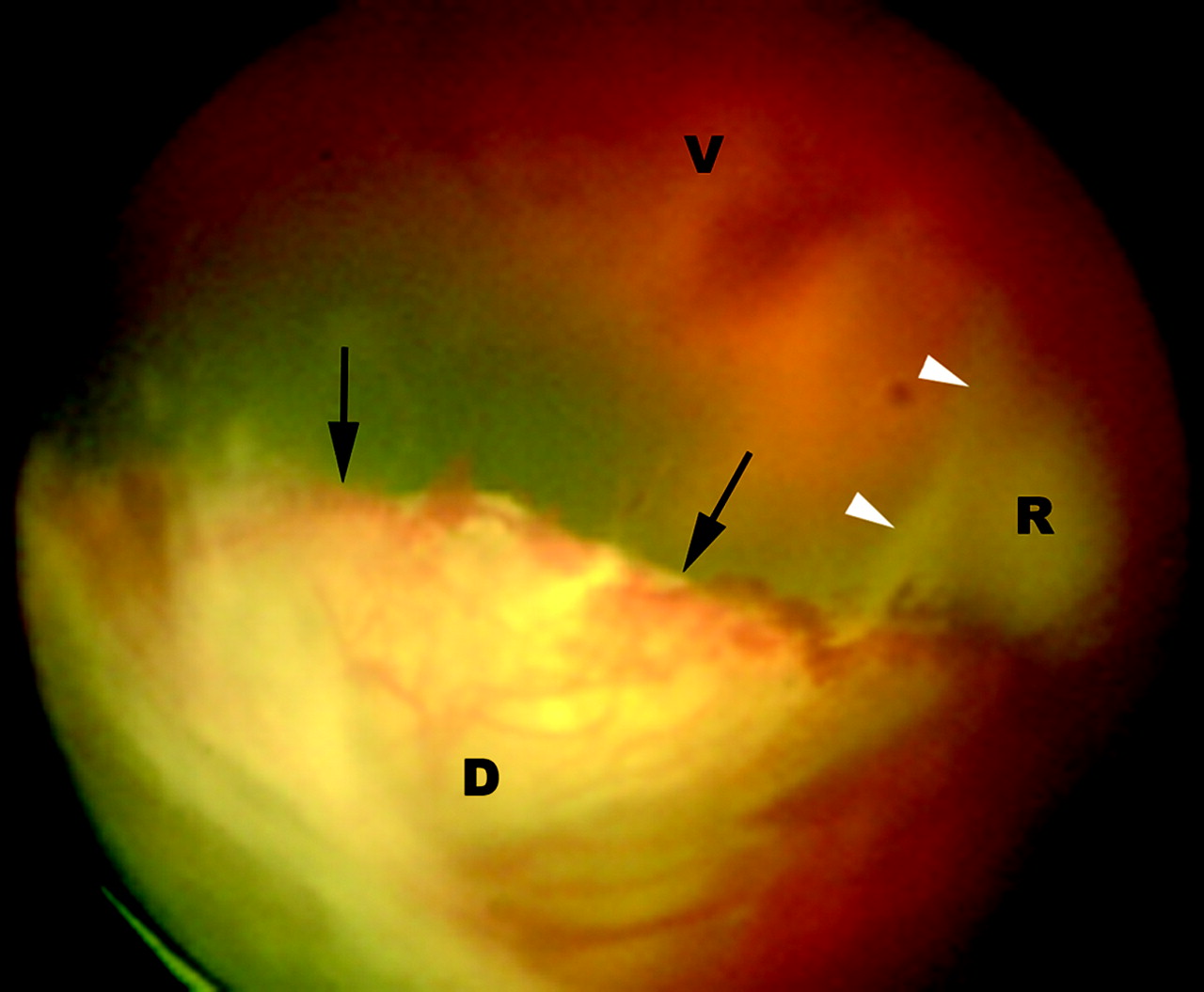
- Fig 1.
Fundus photograph of the left eye shows a retrolental mass (D, arrows) in the inferomedial quadrant of the vitreous (V) with large irregular feeder vessels, focal hemorrhages, and retinal detachment (R, arrowheads).
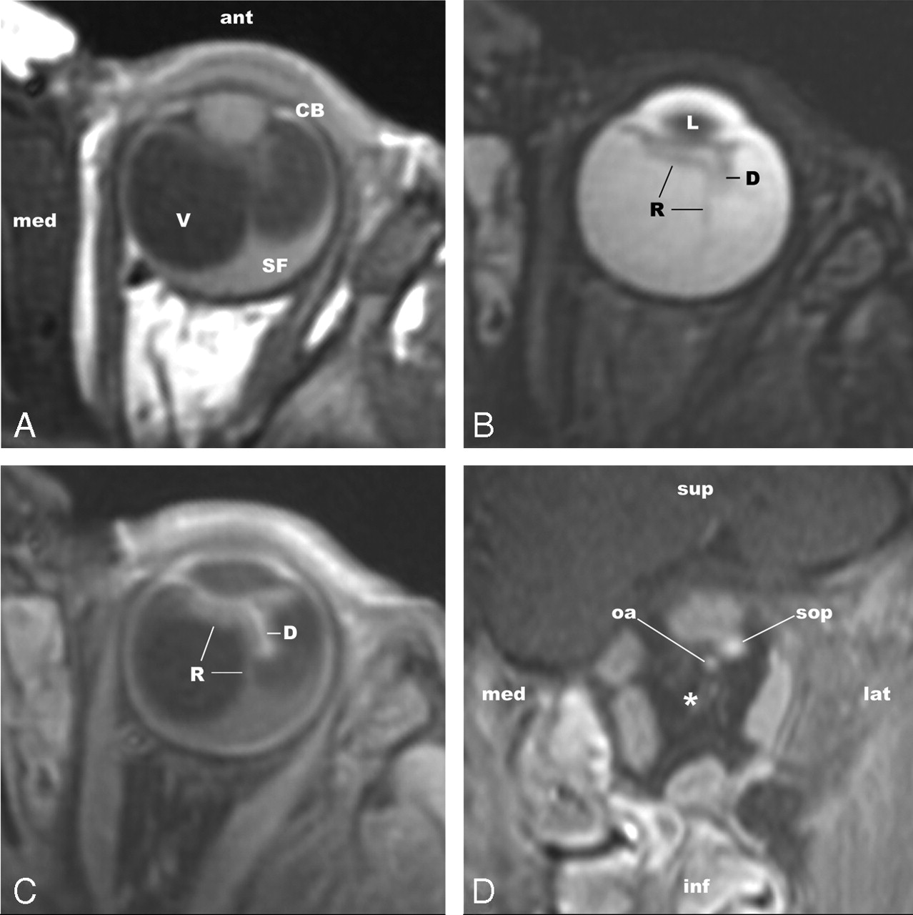
- Fig 2.
Axial T1WI (A) image shows a hyperintense mass in the anterior part of the vitreous (V), adjacent to the ciliary body (CB) on either side of the lens, combined with a tent-shaped retinal detachment with hyperintense subretinal fluid (SF). T2WI (B) image shows retinal detachment as a hypointense fine linear structure (R) and clearly demarcates the dysplastic retinal tissue (D). After we applied the contrast material (C, contrast-enhanced fat-suppressed T1WI image), the latter shows enhancement (D), in contrast to the detached retina (R) and subretinal fluid. Coronal contrast-enhanced fat-suppressed T1WI (D) MR images reveal absence of the optic nerve (asterisk) in an otherwise normal orbit. sop indicates superior ophthalmic vein; oa, ophthalmic artery.
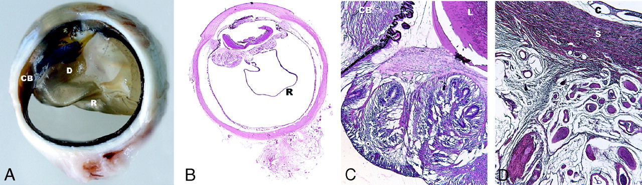
- Fig 3.
A, Gross examination shows the detached retina (R) and the yellowish dysplastic retinal tissue (D), which is adjacent to the ciliary body (CB). The subretinal fluid is located inside the tent-shaped retinal detachment (R) and cannot be seen on this gross specimen. B, Microscopic examination shows the dysplastic retinal tissue behind the lens arising from the retina (R). The mass does not invade the ciliary body or the anterior chamber (hematoxylin-eosin [H&E], original magnification ×3.5 objective). C, Detail of the mass shows still vaguely recognizable retinal tissue near the lens (L) and ciliary body (CB), with rosette formation composed of photoreceptor cells (H&E, original magnification ×20 objective). D, Detail of the region in which the optic nerve was expected shows absence of the optic disc, lamina cribrosa, and optic nerve. Orbital connective tissue with a collection of small (ciliary) vessels is present behind the eye (H&E, original magnification ×20 objective). C indicates choroid; S, sclera.


