
Article Figures & Data
Figures
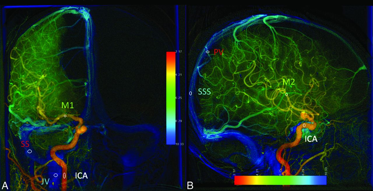
- Fig 1.
Anteroposterior (A) and lateral (B) views of parametric color-coding of quantitative DSA. A, The ROI of the ICA is located at the midpoint of the cervical portion of the ICA. The ROI of M1 is located at the midpoint of the first segment of the middle cerebral artery. The sigmoid sinus ROI is located at the midpoint of the ipsilateral sigmoid sinus. The jugular vein ROI is located in the internal jugular vein at the same level as the ICA ROI. B, The ICA ROI is located in the cavernous portion of the ICA. The M2 ROI is located in the insular branch of the MCA. The PV ROI is located in the outlet of the parietal vein. The SSS ROI is located 2 cm above the confluence of the SSS.
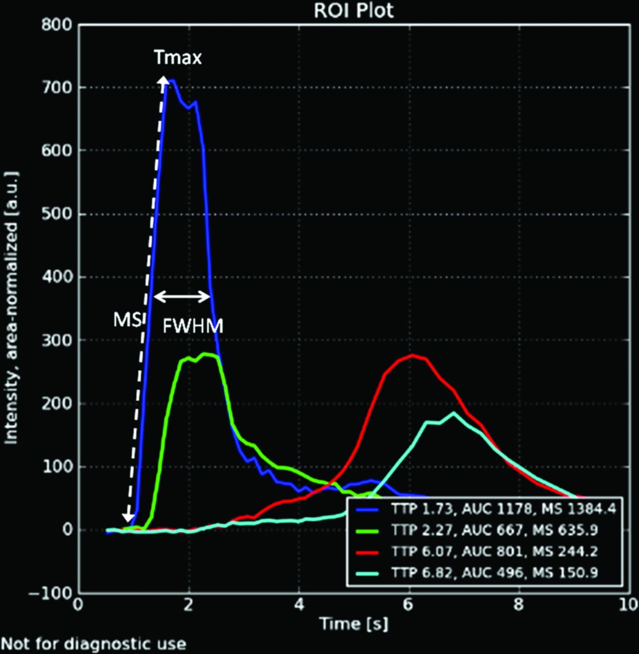
- Fig 2.
Time-attenuation curves of lateral view DSA in a healthy subject. The blue curve represents the TDC of the ROI in the cavernous portion of the ICA. Tmax is the time point at which the ROI reaches maximal intensity. The MS of an ROI is defined by the maximal tangential slope located between arrival time and Tmax. FWHM is the width of the waveform at the level of half maximum concentration.
- Fig 3.
Receiver operating characteristic curves of all significantly different rTmax values (A), all significantly different MS values (B), and all significantly different FWHM values (C) in differentiating patients with stenosis from control groups.
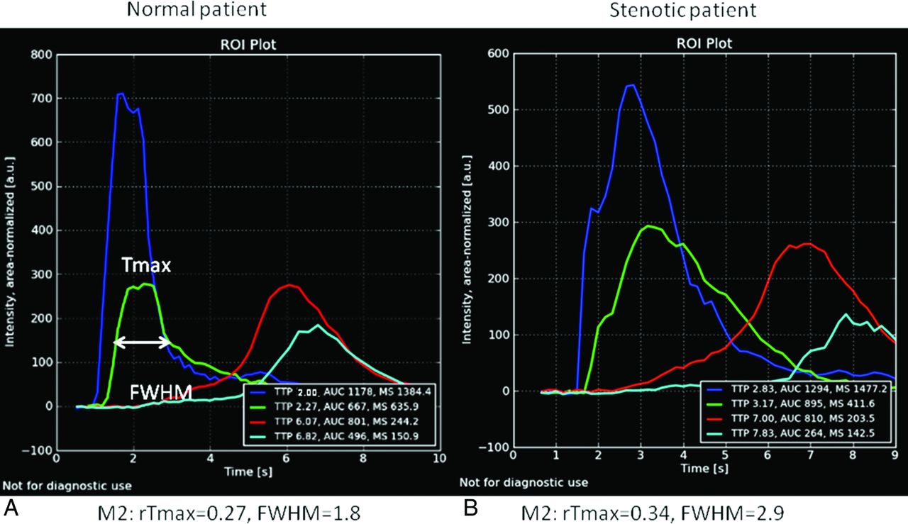
- Fig 4.
TDC of lateral views of angiography are from a healthy individual (A) and a patient with 80% carotid stenosis (B). Both green lines are TDCs of M2. The rTmax of M2 in the patient with stenosis is 0.34 seconds, which was more prolonged than that (0.27 seconds) in the healthy individual. The FWHM of M2 (2.9 seconds) in the patient with stenosis is longer than that (1.8 seconds) in the healthy individual. The ICA (dark blue curves), sigmoid sinus (red curves), and internal jugular vein (light blue curves) all demonstrate prolonged rTmax and are right-shifted in the patient with stenosis. These delayed and dispersed phenomena of angiographic TDCs are consistent with those observed in MR imaging and CT perfusion imaging.


Tables
Group A (Stenosis) Group B (Healthy) P Value No. 70 56 Age (yr) 73.6 ± 11.6 65.6 ± 10.2 <.001a Heart rate (beats/min) 69.9 ± 17.2 72.6 ± 12.3 0.312 Blood pressure (mm Hg) 92.8 ± 19.9 97.6 ± 18.5 0.124 Stenotic degree (%) 81.4% NA NA Prior minor stroke 19 (27%) 1 (1.7%) <.001a Note:—NA indicates not applicable.
↵a Statistically significant (t test, P < .05). There was no measurable stenosis in the healthy (control) population.
ROI Group A Group B P Value Stenosis (n = 70) Healthy (n = 56) rTmax M2 0.77 ± 0.52 0.58 ± 0.32 .005a PV 5.08 ± 1.32 4.38 ± 1.38 .001a SSS 6.35 ± 1.79 5.44 ± 1.44 .001a M1 0.66 ± 0.31 0.46 ± 0.46 .001a SS 6.71 ± 1.91 5.91 ± 1.67 .004a JV 7.29 ± 1.77 6.60 ± 1.62 .008a MS M2 337.15 ± 166.18 390.39 ± 166.98 .03a PV 159.34 ± 73.36 170.56 ± 99.95 .43 SSS 103.78 ± 57.8 134.38 ± 111.07 .03a M1 331.59 ± 144.43 454.44 ± 320.48 .003a SS 100.32 ± 58.22 97.02 ± 47.17 .699 JV 116.78 ± 63.13 136.22 ± 109.63 .185 FWHM M2 2.54 ± 1.14 2.09 ± 0.70 <.001a PV 2.84 ± 2.37 2.74 ± 1.65 .74 SSS 3.49 ± 2.38 3.86 ± 0.75 .607 M1 3.57 ± 1.90 2.78 ± 0.92 .002a SS 4.33 ± 1.83 3.72 ± 1.17 .61 JVb NA NA NA - Table 4:
Cutoff values of 10 significant TDC parameters for detecting stenosis flow with optimized sensitivity and specificity
ROI Variable AUC P Value Cutoff Value Sensitivity Specificity rTmax M2 0.638 (0.555–0.722) .002 0.415 83.6% 37.6% PV 0.615 (0.53–0.701) .011 4.905 55.2% 63.4% SSS 0.655 (0.57–0.74)a .001 6.895 55.2% 73.7% M1 0.663 (0.58–0.746)a .001 0.45 81.0% 58.2% SS 0.582 (0.495–0.669) .045 7.42 73.5% 91.2% JV 0.589 (0.503–0.676) .045 7.77 63.7% 84.9% MS M2 0.606 (0.517–0.695) .02 511.5 89.6% 21.8% M1 0.689 (0.608–0.77)a .041 378.3 66.7% 60.0% SSS 0.636 (0.547–0.725) .002 90.5 55.1% 75.5% FWHM M2 0.609 (0.52–0.699) .013 3.45 57.1% 67.7% M1 0.679 (0.59–0.77)a .001 3.40 57.1% 78.4% Note:—SS indicates sigmoid sinus; JV, internal jugular vein.
↵a The best 4 parameters for detecting stenotic flow.


Jump to section
Related Articles
Cited By...
- Carotid webs produce greater hemodynamic disturbances than atherosclerotic disease: a DSA time-density curve study
- Prolonged cerebral circulation time is more associated with symptomatic carotid stenosis than stenosis degree or collateral circulation
- Peritherapeutic Hemodynamic Changes of Carotid Stenting Evaluated with Quantitative DSA in Patients with Carotid Stenosis