Advertisement

Main menu

  • Home
  • Content
    • Current Issue
    • Accepted Manuscripts
    • Article Preview
    • Past Issue Archive
    • Video Articles
    • AJNR Case Collection
    • Case of the Week Archive
    • Case of the Month Archive
    • Classic Case Archive
  • Special Collections
    • AJNR Awards
    • Low-Field MRI
    • Alzheimer Disease
    • ASNR Foundation Special Collection
    • Photon-Counting CT
    • View All
  • Multimedia
    • AJNR Podcasts
    • AJNR SCANtastic
    • Trainee Corner
    • MRI Safety Corner
    • Imaging Protocols
  • For Authors
    • Submit a Manuscript
    • Submit a Video Article
    • Submit an eLetter to the Editor/Response
    • Manuscript Submission Guidelines
    • Statistical Tips
    • Fast Publishing of Accepted Manuscripts
    • Graphical Abstract Preparation
    • Imaging Protocol Submission
    • Author Policies
  • About Us
    • About AJNR
    • Editorial Board
    • Editorial Board Alumni
  • More
    • Become a Reviewer/Academy of Reviewers
    • Subscribers
    • Permissions
    • Alerts
    • Feedback
    • Advertisers
    • ASNR Home

User menu

  • Alerts
  • Log in

Search

  • Advanced search
American Journal of Neuroradiology

American Journal of Neuroradiology

ASHNR American Society of Functional Neuroradiology ASHNR American Society of Pediatric Neuroradiology ASSR
  • Alerts
  • Log in

Advanced Search

  • Home
  • Content
    • Current Issue
    • Accepted Manuscripts
    • Article Preview
    • Past Issue Archive
    • Video Articles
    • AJNR Case Collection
    • Case of the Week Archive
    • Case of the Month Archive
    • Classic Case Archive
  • Special Collections
    • AJNR Awards
    • Low-Field MRI
    • Alzheimer Disease
    • ASNR Foundation Special Collection
    • Photon-Counting CT
    • View All
  • Multimedia
    • AJNR Podcasts
    • AJNR SCANtastic
    • Trainee Corner
    • MRI Safety Corner
    • Imaging Protocols
  • For Authors
    • Submit a Manuscript
    • Submit a Video Article
    • Submit an eLetter to the Editor/Response
    • Manuscript Submission Guidelines
    • Statistical Tips
    • Fast Publishing of Accepted Manuscripts
    • Graphical Abstract Preparation
    • Imaging Protocol Submission
    • Author Policies
  • About Us
    • About AJNR
    • Editorial Board
    • Editorial Board Alumni
  • More
    • Become a Reviewer/Academy of Reviewers
    • Subscribers
    • Permissions
    • Alerts
    • Feedback
    • Advertisers
    • ASNR Home
  • Follow AJNR on Twitter
  • Visit AJNR on Facebook
  • Follow AJNR on Instagram
  • Join AJNR on LinkedIn
  • RSS Feeds

AJNR Awards, New Junior Editors, and more. Read the latest AJNR updates

Research ArticleAdult Brain

Measuring Glymphatic Flow in Man Using Quantitative Contrast-Enhanced MRI

R. Watts, J.M. Steinklein, L. Waldman, X. Zhou and C.G. Filippi
American Journal of Neuroradiology April 2019, 40 (4) 648-651; DOI: https://doi.org/10.3174/ajnr.A5931
R. Watts
aFrom the Department of Radiology (R.W.), University of Vermont, Burlington, Vermont
  • Find this author on Google Scholar
  • Find this author on PubMed
  • Search for this author on this site
  • ORCID record for R. Watts
J.M. Steinklein
bDepartment of Radiology (J.M.S., L.W., X.Z., C.G.F.), Lenox Hill Hospital and Donald and Barbara Zucker School of Medicine at Hofstra/Northwell, East Garden City, New York.
  • Find this author on Google Scholar
  • Find this author on PubMed
  • Search for this author on this site
  • ORCID record for J.M. Steinklein
L. Waldman
bDepartment of Radiology (J.M.S., L.W., X.Z., C.G.F.), Lenox Hill Hospital and Donald and Barbara Zucker School of Medicine at Hofstra/Northwell, East Garden City, New York.
  • Find this author on Google Scholar
  • Find this author on PubMed
  • Search for this author on this site
  • ORCID record for L. Waldman
X. Zhou
bDepartment of Radiology (J.M.S., L.W., X.Z., C.G.F.), Lenox Hill Hospital and Donald and Barbara Zucker School of Medicine at Hofstra/Northwell, East Garden City, New York.
  • Find this author on Google Scholar
  • Find this author on PubMed
  • Search for this author on this site
  • ORCID record for X. Zhou
C.G. Filippi
bDepartment of Radiology (J.M.S., L.W., X.Z., C.G.F.), Lenox Hill Hospital and Donald and Barbara Zucker School of Medicine at Hofstra/Northwell, East Garden City, New York.
  • Find this author on Google Scholar
  • Find this author on PubMed
  • Search for this author on this site
  • ORCID record for C.G. Filippi
  • Article
  • Figures & Data
  • Info & Metrics
  • Responses
  • References
  • PDF
Loading

Abstract

SUMMARY: On the basis of animal models, glymphatic flow disruption is hypothesized to be a factor in the development of Alzheimer's disease. We report the first quantitative study of glymphatic flow in man, combining intrathecal administration of gadobutrol with serial T1 mapping to produce contrast concentration maps up to 3 days postinjection, demonstrating performing a quantitative study using the techniques described feasibility and providing data on pharmacokinetics.

ABBREVIATION:

AD
Alzheimer's disease

Protein aggregation is the pathologic signature of many neurodegenerative diseases, including Alzheimer's disease (AD).1 The recently discovered glial lymphatic (“glymphatic”) system2 may play a critical role in protein removal, including soluble amyloid-β3 and HPF-τ (Hyperphosphorylated-τ).4 This perivascular glymphatic pathway drives exchange between CSF and interstitial fluid, and dysfunction may be a causal factor in the development of Alzheimer's disease.5 In rodent models, glymphatic flow decreases with age,6 increases during sleep,7 and can be pharmacologically manipulated,7 suggesting similar approaches for patients with AD.

Qualitative MR imaging using percentage signal enhancement on T1-weighted images has been reported in animal models8 and humans.9⇓⇓–12 However, the percentage signal change on a T1-weighted image with contrast concentration depends on the intrinsic T1 of each tissue, the corresponding change in T2*, and the imaging parameters selected. To be able to apply pharmacokinetic models requires consistent determination of contrast concentrations across tissue types and concentrations. Here we use the combination of an intrathecal administration of a gadolinium-based contrast agent (gadobutrol) with quantitative MR imaging to provide maps of contrast concentration versus time, measuring glymphatic flow throughout the brain in a human volunteer at 3T.

Materials and Methods

Patient History

A 55-year-old man referred for MR myelography was recruited for this Health Insurance Portability and Accountability Act–compliant, institutional review board (The Feinstein Institute for Medical Research)–approved study using an intrathecal injection of 0.5 mL of 1.0 mmol of gadobutrol. The patient gave written, informed consent.

Data Acquisition and Analysis

Quantitative T1-mapping data were acquired on a 3T Achieva TX MR imaging scanner (Philips Healthcare, Best, the Netherlands) with an 8-channel head coil using a multiple flip angle 3D spoiled gradient-echo sequence with TE/TR = 2.8/20 ms; flip angles = 2°, 5°, 10°, 20°, 40°; FOV = 240 × 228 × 120 mm3; acquired resolution = 1.2 × 1.2 × 1.2 mm3. Acquisition time was 3 minutes 47 seconds for each flip angle using a sensitivity encoding factor of 2, to give a total time of 24 minutes. Scans were acquired at baseline and at 9 further acquisitions during the first 10 hours (60, 210, 240, 270, 370, 450, 500, 560, and 620 minutes), followed by acquisitions at 26, 50, and 79 hours. The subject was not restricted but was asked to remain supine for as much as possible during the first 10 hours but then was allowed to move normally for the remaining time. He had a normal night's sleep before being scanned the final 3 times.

Data Analysis: Theory

The method is based on the nuclear MR steady-state signal equation, Embedded Image where E1 = Embedded Image, Si is the signal intensity for each flip angle αi, and M0 is a constant representing the equilibrium magnetization. This equation can be transformed to a linear form, from which the gradient E1 and hence the tissue T1 can be determined.13

The T1 value is related to the contrast agent concentration by Embedded Image Where T10 is the native T1 value of the tissue (s), and r1 is the relaxivity of the contrast agent, assumed to be 5.1 l/mmol/s for gadobutrol.

Data Processing

Each volume was coregistered to the 20° flip angle acquisition from the first scan acquired before contrast injection using SPM12 (http://www.fil.ion.ucl.ac.uk/spm/software/spm12). T1 maps were generated using least squares fitting to the signal equation using custom Matlab (MathWorks, Natick, Massachusetts) scripts. The precontrast map provided the baseline T1 values (T10), enabling contrast concentration to be calculated at each subsequent time.

Brain segmentation was performed using SPM12 on the 20° flip angle acquisition from the first scan acquired before contrast injection, resulting in native space gray matter, white matter, and CSF components. Cortical gray matter masks were defined by the intersection of the gray matter mask and the corresponding regions of the Montreal Neurological Institute structural atlas transformed to native space. Juxtacortical white matter and CSF masks were defined by the intersection of their native tissue components with the cortical gray matter mask expanded by 3 mm using only voxels with >95% probability of being the target tissue.

Results

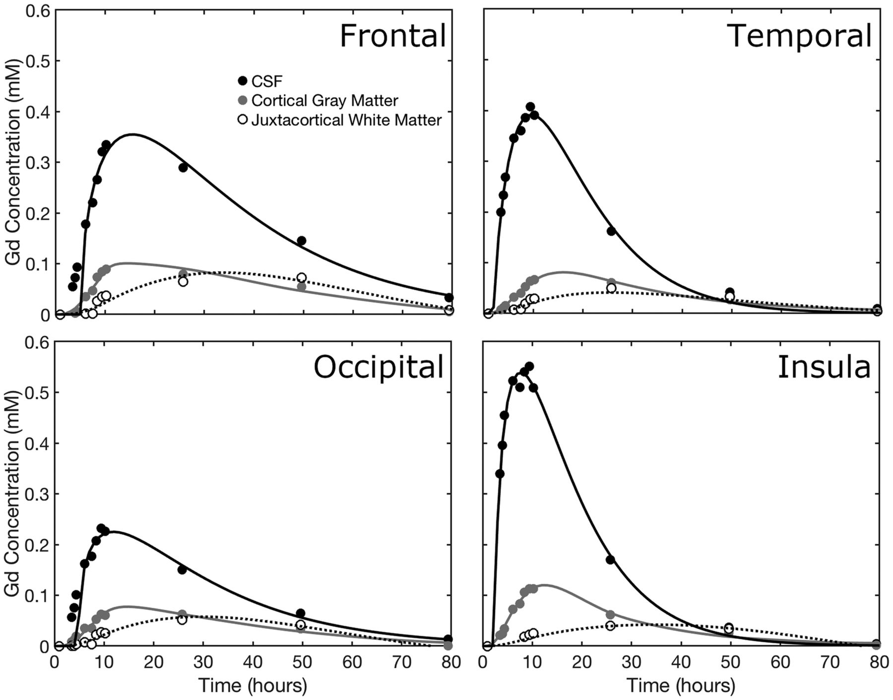
The time course of contrast concentration during 79 hours postinjection is shown in Fig 1. Contrast arrived in the cisterna magna between 1 and 3 hours postinjection and covered the entire subarachnoid space at 8 hours. The subarachnoid CSF concentration peaked between 10 and 15 hours at approximately 0.5 mmol/L. Cortical enhancement measurable from 4 hours peaked between 10 and 26 hours, with a maximum concentration of approximately 0.1 mmol/L. The largest concentration was noted in the temporal lobes and insula in the first 10 hours. Cortical contrast remained at 50 hours with near-complete clearance at 79 hours. Depending on location, white matter concentration peaked between 26 and 50 hours.

Fig 1.
  • Download figure
  • Open in new tab
  • Download powerpoint
Fig 1.

Quantitative maps of gadolinium tracer across time. The tracer arrived in the cisterna magna between 1 and 3.5 hours after intrathecal injection. Superior parts of the subarachnoid space enhance late, with clearance also delayed.

Concentration-time curves were calculated for cortical gray matter and juxtacortical white matter (Fig 2). Biologic half-life of gadobutrol in the subarachnoid CSF, based on the washout curve, was approximately 12 hours. A 2-compartment model provided a good fit to the cortical contrast concentration curve.

Fig 2.
  • Download figure
  • Open in new tab
  • Download powerpoint
Fig 2.

The time course of gadolinium concentration for the frontal, temporal, occipital, and parietal lobes. CSF concentration, cortical gray matter, and juxtacortical white matter curves are shown. Juxtacortical white matter is within 3 mm of the cortex. In each case, the CSF tracer concentration is fitted to a γ-variate distribution, while the tissue concentrations are fitted by 2-compartment pharmacokinetic models.

Discussion

Most current literature on glymphatic flow is based on rodent models using highly invasive techniques. A few studies used MR imaging qualitatively to investigate glymphatic flow in rodents8 and humans.9⇓⇓–12 Our report improves on those studies by quantifying contrast concentration. We are only aware of a single study that has used similar quantification in rats at 9.4T.14 Compared with rodents, contrast uptake and clearance are much slower in our subject but are still faster than would be expected due to only passive diffusion, which provides further evidence that intrathecal administration of drugs may provide access to the entire brain parenchyma.

We observed minimal enhancement of the lateral ventricles at any time, suggesting that contrast entering the ventricular system is rapidly diluted.

Although only a single individual was imaged, the acquisition of serial quantitative data at many time points during 3 days represents a unique dataset. While cost and inconvenience to patients of an identical approach are prohibitive for routine use, our results suggest that an abbreviated protocol may suffice.

Regarding retention of gadolinium-based contrast agents, recent studies including long-term follow-up after intrathecal contrast injection have not identified adverse effects.15,16 Gadolinium retention after intrathecal administration was not observed for gadobutrol at the same dose used in the current study.11

Conclusions

We have demonstrated the feasibility of using T1 mapping to quantify contrast concentration to analyze glymphatic flow in man, for which there is increasing interest in its use as a biomarker and potential therapeutic target in AD.

Footnotes

  • Disclosures: Richard Watts—RELATED: Grant: Foundation of the American Society of Neuroradiology Boerger Research Fund for Alzheimer's Disease and Neurocognitive Disorders.* Christopher G. Filippi—RELATED: Grant: Foundation of the American Society of Neuroradiology, Comments: Grant money was given by the Foundation of the American Society of Neuroradiology Boerger Fund to support this research study*; UNRELATED: Consultancy: Syntactx Inc, Comments: I read MR imaging examinations as part of clinical trials work. *Money paid to the institution.

  • This study was funded by the Foundation of the American Society of Neuroradiology Boerger Research Fund for Alzheimer's Disease and Neurocognitive Disorders.

  • Paper previously presented, in part, at: Annual Meeting of the American Society of Functional Neuroradiology, October 15–17, 2018; Coronado, California.

REFERENCES

  1. 1.↵
    1. Ross CA,
    2. Poirier MA
    . Protein aggregation and neurodegenerative disease. Nat Med 2004;10(Suppl):S10–17 pmid:15272267
    CrossRefPubMed
  2. 2.↵
    1. Iliff JJ,
    2. Wang M,
    3. Liao Y, et al
    . A paravascular pathway facilitates CSF flow through the brain parenchyma and the clearance of interstitial solutes, including amyloid β. Sci Transl Med 2012;4:147ra111 doi:10.1126/scitranslmed.3003748 pmid:22896675
    Abstract/FREE Full Text
  3. 3.↵
    1. Hawkes CA,
    2. Sullivan PM,
    3. Hands S, et al
    . Disruption of arterial perivascular drainage of amyloid-β from the brains of mice expressing the human APOE ε4 allele. PLoS One 2012;7:e41636 doi:10.1371/journal.pone.0041636 pmid:22848551
    CrossRefPubMed
  4. 4.↵
    1. Iliff JJ,
    2. Chen MJ,
    3. Plog BA, et al
    . Impairment of glymphatic pathway function promotes tau pathology after traumatic brain injury. J Neurosci 2014;34:16180–93 doi:10.1523/JNEUROSCI.3020-14.2014 pmid:25471560
    Abstract/FREE Full Text
  5. 5.↵
    1. Bakker EN,
    2. Bacskai BJ,
    3. Arbel-Ornath M, et al
    . Lymphatic clearance of the brain: perivascular, paravascular and significance for neurodegenerative diseases. Cell Mol Neurobiol 2016;36:181–94 doi:10.1007/s10571-015-0273-8 pmid:26993512
    CrossRefPubMed
  6. 6.↵
    1. Hawkes CA,
    2. Hartig W,
    3. Kacza J, et al
    . Perivascular drainage of solutes is impaired in the ageing mouse brain and in the presence of cerebral amyloid angiopathy. Acta Neuropathol 2011;121:431–43 doi:10.1007/s00401-011-0801-7 pmid:21259015
    CrossRefPubMed
  7. 7.↵
    1. Xie L,
    2. Kang H,
    3. Xu Q, et al
    . Sleep drives metabolite clearance from the adult brain. Science 2013;342:373–77 doi:10.1126/science.1241224 pmid:24136970
    Abstract/FREE Full Text
  8. 8.↵
    1. Iliff JJ,
    2. Lee H,
    3. Yu M, et al
    . Brain-wide pathway for waste clearance captured by contrast-enhanced MRI. J Clin Invest 2013;123:1299–309 doi:10.1172/JCI67677 pmid:23434588
    CrossRefPubMed
  9. 9.↵
    1. Eide PK,
    2. Ringstad G
    . MRI with intrathecal MRI gadolinium contrast medium administration: a possible method to assess glymphatic function in human brain. Acta Radiol Open 2015;4:2058460115609635 doi:10.1177/2058460115609635 pmid:26634147
    CrossRefPubMed
  10. 10.↵
    1. Eide PK,
    2. Ringstad G
    . Delayed clearance of cerebrospinal fluid tracer from entorhinal cortex in idiopathic normal pressure hydrocephalus: a glymphatic magnetic resonance imaging study. J Cereb Blood Flow Metab 2018; Feb 27:271678X18760974. [Epub ahead of print] doi:10.1177/0271678X18760974 pmid:29485341
    CrossRefPubMed
  11. 11.↵
    1. Ringstad G,
    2. Valnes LM,
    3. Dale AM, et al
    . Brain-wide glymphatic enhancement and clearance in humans assessed with MRI. JCI Insight 2018 Jul 12;3(13). [Epub ahead of print] doi:10.1172/jci.insight.121537 pmid:29997300
    CrossRefPubMed
  12. 12.↵
    1. Ringstad G,
    2. Vatnehol SAS,
    3. Eide PK
    . Glymphatic MRI in idiopathic normal pressure hydrocephalus. Brain 2017;140:2691–705 doi:10.1093/brain/awx191 pmid:28969373
    CrossRefPubMed
  13. 13.↵
    1. Cheng HL,
    2. Wright GA
    . Rapid high-resolution T(1) mapping by variable flip angles: accurate and precise measurements in the presence of radiofrequency field inhomogeneity. Magnetic Reson Med 2006;55:566–74 doi:10.1002/mrm.20791 pmid:16450365
    CrossRefPubMed
  14. 14.↵
    1. Lee H,
    2. Mortensen K,
    3. Sanggaard S, et al
    . Quantitative Gd-DOTA uptake from cerebrospinal fluid into rat brain using 3D VFA-SPGR at 9.4T. Magnetic Reson Med 2018;79:1568–78 doi:10.1002/mrm.26779 pmid:28627037
    CrossRefPubMed
  15. 15.↵
    1. Safriel Y,
    2. Ang R,
    3. Ali M
    . Gadolinium use in spine pain management procedures for patients with contrast allergies: results in 527 procedures. Cardiovasc Intervent Radiol 2008;31:325–31 doi:10.1007/s00270-007-9192-5 pmid:17939000
    CrossRefPubMed
  16. 16.↵
    1. Nacar Dogan S,
    2. Kizilkilic O,
    3. Kocak B, et al
    . Intrathecal gadolinium-enhanced MR cisternography in patients with otorhinorrhea: 10-year experience of a tertiary referral center. Neuroradiology 2018;60:471–77 doi:10.1007/s00234-018-2014-4 pmid:29572604
    CrossRefPubMed
  • Received August 22, 2018.
  • Accepted after revision November 13, 2018.
  • © 2019 by American Journal of Neuroradiology
View Abstract
PreviousNext
Back to top

In this issue

American Journal of Neuroradiology: 40 (4)
American Journal of Neuroradiology
Vol. 40, Issue 4
1 Apr 2019
  • Table of Contents
  • Index by author
  • Complete Issue (PDF)
Advertisement
Print
Download PDF
Email Article

Thank you for your interest in spreading the word on American Journal of Neuroradiology.

NOTE: We only request your email address so that the person you are recommending the page to knows that you wanted them to see it, and that it is not junk mail. We do not capture any email address.

Enter multiple addresses on separate lines or separate them with commas.
Measuring Glymphatic Flow in Man Using Quantitative Contrast-Enhanced MRI
(Your Name) has sent you a message from American Journal of Neuroradiology
(Your Name) thought you would like to see the American Journal of Neuroradiology web site.
CAPTCHA
This question is for testing whether or not you are a human visitor and to prevent automated spam submissions.
Cite this article
R. Watts, J.M. Steinklein, L. Waldman, X. Zhou, C.G. Filippi
Measuring Glymphatic Flow in Man Using Quantitative Contrast-Enhanced MRI
American Journal of Neuroradiology Apr 2019, 40 (4) 648-651; DOI: 10.3174/ajnr.A5931

Citation Manager Formats

  • BibTeX
  • Bookends
  • EasyBib
  • EndNote (tagged)
  • EndNote 8 (xml)
  • Medlars
  • Mendeley
  • Papers
  • RefWorks Tagged
  • Ref Manager
  • RIS
  • Zotero
0 Responses
Respond to this article
Share
Bookmark this article
Measuring Glymphatic Flow in Man Using Quantitative Contrast-Enhanced MRI
R. Watts, J.M. Steinklein, L. Waldman, X. Zhou, C.G. Filippi
American Journal of Neuroradiology Apr 2019, 40 (4) 648-651; DOI: 10.3174/ajnr.A5931
del.icio.us logo Twitter logo Facebook logo Mendeley logo
  • Tweet Widget
  • Facebook Like
  • Google Plus One
Purchase

Jump to section

  • Article
    • Abstract
    • ABBREVIATION:
    • Materials and Methods
    • Results
    • Discussion
    • Conclusions
    • Footnotes
    • REFERENCES
  • Figures & Data
  • Info & Metrics
  • Responses
  • References
  • PDF

Related Articles

  • No related articles found.
  • PubMed
  • Google Scholar

Cited By...

  • Heterogeneity in human brain clearance adds resilience against tauopathy - a computational model informed by glymphatic MRI
  • In-silico molecular enrichment and clearance of the human intracranial space
  • Different Glymphatic Kinetics in Spontaneous Intracranial Hypotension
  • Glymphatic dysfunction in multiple sclerosis and its association with disease pathology and disability
  • Preliminary cross-sectional investigations into the human glymphatic system using multiple novel non-contrast MRI methods
  • Human brain solute transport quantified by glymphatic MRI-informed biophysics during sleep and sleep deprivation
  • Intrathecal [64Cu]Cu-albumin PET reveals age-related decline of cerebrospinal fluid (CSF)-lymphatic efflux
  • CSF circulation and dispersion yield rapid clearance from intracranial compartments
  • Association of Sleep, Neuropsychological Performance, and Gray Matter Volume With Glymphatic Function in Community-Dwelling Older Adults
  • Intrathecal Use of Gadobutrol for Glymphatic MR Imaging: Prospective Safety Study of 100 Patients
  • Crossref
  • Google Scholar

This article has not yet been cited by articles in journals that are participating in Crossref Cited-by Linking.

More in this TOC Section

  • Diagnostic Neuroradiology of Monoclonal Antibodies
  • Clinical Outcomes After Chiari I Decompression
  • Segmentation of Brain Metastases with BLAST
Show more Adult Brain

Similar Articles

Advertisement

Indexed Content

  • Current Issue
  • Accepted Manuscripts
  • Article Preview
  • Past Issues
  • Editorials
  • Editor's Choice
  • Fellows' Journal Club
  • Letters to the Editor
  • Video Articles

Cases

  • Case Collection
  • Archive - Case of the Week
  • Archive - Case of the Month
  • Archive - Classic Case

More from AJNR

  • Trainee Corner
  • Imaging Protocols
  • MRI Safety Corner
  • Book Reviews

Multimedia

  • AJNR Podcasts
  • AJNR Scantastics

Resources

  • Turnaround Time
  • Submit a Manuscript
  • Submit a Video Article
  • Submit an eLetter to the Editor/Response
  • Manuscript Submission Guidelines
  • Statistical Tips
  • Fast Publishing of Accepted Manuscripts
  • Graphical Abstract Preparation
  • Imaging Protocol Submission
  • Evidence-Based Medicine Level Guide
  • Publishing Checklists
  • Author Policies
  • Become a Reviewer/Academy of Reviewers
  • News and Updates

About Us

  • About AJNR
  • Editorial Board
  • Editorial Board Alumni
  • Alerts
  • Permissions
  • Not an AJNR Subscriber? Join Now
  • Advertise with Us
  • Librarian Resources
  • Feedback
  • Terms and Conditions
  • AJNR Editorial Board Alumni

American Society of Neuroradiology

  • Not an ASNR Member? Join Now

© 2025 by the American Society of Neuroradiology All rights, including for text and data mining, AI training, and similar technologies, are reserved.
Print ISSN: 0195-6108 Online ISSN: 1936-959X

Powered by HighWire