Advertisement

Main menu

  • Home
  • Content
    • Current Issue
    • Accepted Manuscripts
    • Article Preview
    • Past Issue Archive
    • Video Articles
    • AJNR Case Collection
    • Case of the Week Archive
    • Case of the Month Archive
    • Classic Case Archive
  • Special Collections
    • AJNR Awards
    • Low-Field MRI
    • Alzheimer Disease
    • ASNR Foundation Special Collection
    • Photon-Counting CT
    • View All
  • Multimedia
    • AJNR Podcasts
    • AJNR SCANtastic
    • Trainee Corner
    • MRI Safety Corner
    • Imaging Protocols
  • For Authors
    • Submit a Manuscript
    • Submit a Video Article
    • Submit an eLetter to the Editor/Response
    • Manuscript Submission Guidelines
    • Statistical Tips
    • Fast Publishing of Accepted Manuscripts
    • Graphical Abstract Preparation
    • Imaging Protocol Submission
    • Author Policies
  • About Us
    • About AJNR
    • Editorial Board
    • Editorial Board Alumni
  • More
    • Become a Reviewer/Academy of Reviewers
    • Subscribers
    • Permissions
    • Alerts
    • Feedback
    • Advertisers
    • ASNR Home

User menu

  • Alerts
  • Log in

Search

  • Advanced search
American Journal of Neuroradiology

American Journal of Neuroradiology

ASHNR American Society of Functional Neuroradiology ASHNR American Society of Pediatric Neuroradiology ASSR
  • Alerts
  • Log in

Advanced Search

  • Home
  • Content
    • Current Issue
    • Accepted Manuscripts
    • Article Preview
    • Past Issue Archive
    • Video Articles
    • AJNR Case Collection
    • Case of the Week Archive
    • Case of the Month Archive
    • Classic Case Archive
  • Special Collections
    • AJNR Awards
    • Low-Field MRI
    • Alzheimer Disease
    • ASNR Foundation Special Collection
    • Photon-Counting CT
    • View All
  • Multimedia
    • AJNR Podcasts
    • AJNR SCANtastic
    • Trainee Corner
    • MRI Safety Corner
    • Imaging Protocols
  • For Authors
    • Submit a Manuscript
    • Submit a Video Article
    • Submit an eLetter to the Editor/Response
    • Manuscript Submission Guidelines
    • Statistical Tips
    • Fast Publishing of Accepted Manuscripts
    • Graphical Abstract Preparation
    • Imaging Protocol Submission
    • Author Policies
  • About Us
    • About AJNR
    • Editorial Board
    • Editorial Board Alumni
  • More
    • Become a Reviewer/Academy of Reviewers
    • Subscribers
    • Permissions
    • Alerts
    • Feedback
    • Advertisers
    • ASNR Home
  • Follow AJNR on Twitter
  • Visit AJNR on Facebook
  • Follow AJNR on Instagram
  • Join AJNR on LinkedIn
  • RSS Feeds

AJNR Awards, New Junior Editors, and more. Read the latest AJNR updates

Research ArticleAdult Brain

Measuring Glymphatic Flow in Man Using Quantitative Contrast-Enhanced MRI

R. Watts, J.M. Steinklein, L. Waldman, X. Zhou and C.G. Filippi
American Journal of Neuroradiology April 2019, 40 (4) 648-651; DOI: https://doi.org/10.3174/ajnr.A5931
R. Watts
aFrom the Department of Radiology (R.W.), University of Vermont, Burlington, Vermont
  • Find this author on Google Scholar
  • Find this author on PubMed
  • Search for this author on this site
  • ORCID record for R. Watts
J.M. Steinklein
bDepartment of Radiology (J.M.S., L.W., X.Z., C.G.F.), Lenox Hill Hospital and Donald and Barbara Zucker School of Medicine at Hofstra/Northwell, East Garden City, New York.
  • Find this author on Google Scholar
  • Find this author on PubMed
  • Search for this author on this site
  • ORCID record for J.M. Steinklein
L. Waldman
bDepartment of Radiology (J.M.S., L.W., X.Z., C.G.F.), Lenox Hill Hospital and Donald and Barbara Zucker School of Medicine at Hofstra/Northwell, East Garden City, New York.
  • Find this author on Google Scholar
  • Find this author on PubMed
  • Search for this author on this site
  • ORCID record for L. Waldman
X. Zhou
bDepartment of Radiology (J.M.S., L.W., X.Z., C.G.F.), Lenox Hill Hospital and Donald and Barbara Zucker School of Medicine at Hofstra/Northwell, East Garden City, New York.
  • Find this author on Google Scholar
  • Find this author on PubMed
  • Search for this author on this site
  • ORCID record for X. Zhou
C.G. Filippi
bDepartment of Radiology (J.M.S., L.W., X.Z., C.G.F.), Lenox Hill Hospital and Donald and Barbara Zucker School of Medicine at Hofstra/Northwell, East Garden City, New York.
  • Find this author on Google Scholar
  • Find this author on PubMed
  • Search for this author on this site
  • ORCID record for C.G. Filippi
  • Article
  • Figures & Data
  • Info & Metrics
  • Responses
  • References
  • PDF
Loading

Article Figures & Data

Figures

  • Fig 1.
    • Download figure
    • Open in new tab
    • Download powerpoint
    Fig 1.

    Quantitative maps of gadolinium tracer across time. The tracer arrived in the cisterna magna between 1 and 3.5 hours after intrathecal injection. Superior parts of the subarachnoid space enhance late, with clearance also delayed.
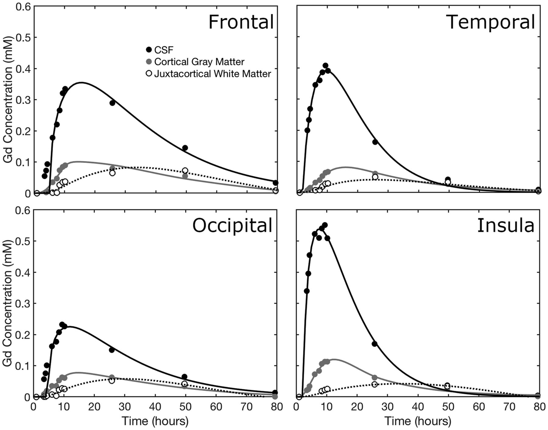
  • Fig 2.
    • Download figure
    • Open in new tab
    • Download powerpoint
    Fig 2.

    The time course of gadolinium concentration for the frontal, temporal, occipital, and parietal lobes. CSF concentration, cortical gray matter, and juxtacortical white matter curves are shown. Juxtacortical white matter is within 3 mm of the cortex. In each case, the CSF tracer concentration is fitted to a γ-variate distribution, while the tissue concentrations are fitted by 2-compartment pharmacokinetic models.

PreviousNext
Back to top

In this issue

American Journal of Neuroradiology: 40 (4)
American Journal of Neuroradiology
Vol. 40, Issue 4
1 Apr 2019
  • Table of Contents
  • Index by author
  • Complete Issue (PDF)
Advertisement
Print
Download PDF
Email Article

Thank you for your interest in spreading the word on American Journal of Neuroradiology.

NOTE: We only request your email address so that the person you are recommending the page to knows that you wanted them to see it, and that it is not junk mail. We do not capture any email address.

Enter multiple addresses on separate lines or separate them with commas.
Measuring Glymphatic Flow in Man Using Quantitative Contrast-Enhanced MRI
(Your Name) has sent you a message from American Journal of Neuroradiology
(Your Name) thought you would like to see the American Journal of Neuroradiology web site.
CAPTCHA
This question is for testing whether or not you are a human visitor and to prevent automated spam submissions.
Cite this article
R. Watts, J.M. Steinklein, L. Waldman, X. Zhou, C.G. Filippi
Measuring Glymphatic Flow in Man Using Quantitative Contrast-Enhanced MRI
American Journal of Neuroradiology Apr 2019, 40 (4) 648-651; DOI: 10.3174/ajnr.A5931

Citation Manager Formats

  • BibTeX
  • Bookends
  • EasyBib
  • EndNote (tagged)
  • EndNote 8 (xml)
  • Medlars
  • Mendeley
  • Papers
  • RefWorks Tagged
  • Ref Manager
  • RIS
  • Zotero
0 Responses
Respond to this article
Share
Bookmark this article
Measuring Glymphatic Flow in Man Using Quantitative Contrast-Enhanced MRI
R. Watts, J.M. Steinklein, L. Waldman, X. Zhou, C.G. Filippi
American Journal of Neuroradiology Apr 2019, 40 (4) 648-651; DOI: 10.3174/ajnr.A5931
del.icio.us logo Twitter logo Facebook logo Mendeley logo
  • Tweet Widget
  • Facebook Like
  • Google Plus One
Purchase

Jump to section

  • Article
    • Abstract
    • ABBREVIATION:
    • Materials and Methods
    • Results
    • Discussion
    • Conclusions
    • Footnotes
    • REFERENCES
  • Figures & Data
  • Info & Metrics
  • Responses
  • References
  • PDF

Related Articles

  • No related articles found.
  • PubMed
  • Google Scholar

Cited By...

  • T2-weighted T1 mapping and automated segmentation of CSF: Assessment of solute gradients in the healthy brain
  • Heterogeneity in human brain clearance adds resilience against tauopathy - a computational model informed by glymphatic MRI
  • In-silico molecular enrichment and clearance of the human intracranial space
  • Different Glymphatic Kinetics in Spontaneous Intracranial Hypotension
  • Glymphatic dysfunction in multiple sclerosis and its association with disease pathology and disability
  • Preliminary cross-sectional investigations into the human glymphatic system using multiple novel non-contrast MRI methods
  • Human brain solute transport quantified by glymphatic MRI-informed biophysics during sleep and sleep deprivation
  • Intrathecal [64Cu]Cu-albumin PET reveals age-related decline of cerebrospinal fluid (CSF)-lymphatic efflux
  • CSF circulation and dispersion yield rapid clearance from intracranial compartments
  • Association of Sleep, Neuropsychological Performance, and Gray Matter Volume With Glymphatic Function in Community-Dwelling Older Adults
  • Intrathecal Use of Gadobutrol for Glymphatic MR Imaging: Prospective Safety Study of 100 Patients
  • Crossref
  • Google Scholar

This article has not yet been cited by articles in journals that are participating in Crossref Cited-by Linking.

More in this TOC Section

  • Diagnostic Neuroradiology of Monoclonal Antibodies
  • Clinical Outcomes After Chiari I Decompression
  • Segmentation of Brain Metastases with BLAST
Show more Adult Brain

Similar Articles

Advertisement

Indexed Content

  • Current Issue
  • Accepted Manuscripts
  • Article Preview
  • Past Issues
  • Editorials
  • Editor's Choice
  • Fellows' Journal Club
  • Letters to the Editor
  • Video Articles

Cases

  • Case Collection
  • Archive - Case of the Week
  • Archive - Case of the Month
  • Archive - Classic Case

More from AJNR

  • Trainee Corner
  • Imaging Protocols
  • MRI Safety Corner
  • Book Reviews

Multimedia

  • AJNR Podcasts
  • AJNR Scantastics

Resources

  • Turnaround Time
  • Submit a Manuscript
  • Submit a Video Article
  • Submit an eLetter to the Editor/Response
  • Manuscript Submission Guidelines
  • Statistical Tips
  • Fast Publishing of Accepted Manuscripts
  • Graphical Abstract Preparation
  • Imaging Protocol Submission
  • Evidence-Based Medicine Level Guide
  • Publishing Checklists
  • Author Policies
  • Become a Reviewer/Academy of Reviewers
  • News and Updates

About Us

  • About AJNR
  • Editorial Board
  • Editorial Board Alumni
  • Alerts
  • Permissions
  • Not an AJNR Subscriber? Join Now
  • Advertise with Us
  • Librarian Resources
  • Feedback
  • Terms and Conditions
  • AJNR Editorial Board Alumni

American Society of Neuroradiology

  • Not an ASNR Member? Join Now

© 2025 by the American Society of Neuroradiology All rights, including for text and data mining, AI training, and similar technologies, are reserved.
Print ISSN: 0195-6108 Online ISSN: 1936-959X

Powered by HighWire