Advertisement

Main menu

  • Home
  • Content
    • Current Issue
    • Accepted Manuscripts
    • Article Preview
    • Past Issue Archive
    • AJNR Case Collection
    • Case of the Week Archive
    • Classic Case Archive
    • Case of the Month Archive
  • Special Collections
    • Spinal CSF Leak Articles (Jan 2020-June 2024)
    • 2024 AJNR Journal Awards
    • Most Impactful AJNR Articles
  • Multimedia
    • AJNR Podcast
    • AJNR Scantastics
    • Video Articles
  • For Authors
    • Submit a Manuscript
    • Author Policies
    • Fast publishing of Accepted Manuscripts
    • Graphical Abstract Preparation
    • Manuscript Submission Guidelines
    • Imaging Protocol Submission
    • Submit a Case for the Case Collection
  • About Us
    • About AJNR
    • Editorial Board
  • More
    • Become a Reviewer/Academy of Reviewers
    • Subscribers
    • Permissions
    • Alerts
    • Feedback
    • Advertisers
    • ASNR Home
  • Other Publications
    • ajnr

User menu

  • Alerts
  • Log in

Search

  • Advanced search
American Journal of Neuroradiology

American Journal of Neuroradiology

ASHNR American Society of Functional Neuroradiology ASHNR American Society of Pediatric Neuroradiology ASSR
  • Alerts
  • Log in

Advanced Search

  • Home
  • Content
    • Current Issue
    • Accepted Manuscripts
    • Article Preview
    • Past Issue Archive
    • AJNR Case Collection
    • Case of the Week Archive
    • Classic Case Archive
    • Case of the Month Archive
  • Special Collections
    • Spinal CSF Leak Articles (Jan 2020-June 2024)
    • 2024 AJNR Journal Awards
    • Most Impactful AJNR Articles
  • Multimedia
    • AJNR Podcast
    • AJNR Scantastics
    • Video Articles
  • For Authors
    • Submit a Manuscript
    • Author Policies
    • Fast publishing of Accepted Manuscripts
    • Graphical Abstract Preparation
    • Manuscript Submission Guidelines
    • Imaging Protocol Submission
    • Submit a Case for the Case Collection
  • About Us
    • About AJNR
    • Editorial Board
  • More
    • Become a Reviewer/Academy of Reviewers
    • Subscribers
    • Permissions
    • Alerts
    • Feedback
    • Advertisers
    • ASNR Home
  • Follow AJNR on Twitter
  • Visit AJNR on Facebook
  • Follow AJNR on Instagram
  • Join AJNR on LinkedIn
  • RSS Feeds

Welcome to the new AJNR, Updated Hall of Fame, and more. Read the full announcements.


AJNR is seeking candidates for the position of Associate Section Editor, AJNR Case Collection. Read the full announcement.


Research ArticleHead & Neck
Open Access

Reduced Jet Velocity in Venous Flow after CSF Drainage: Assessing Hemodynamic Causes of Pulsatile Tinnitus

H. Haraldsson, J.R. Leach, E.I. Kao, A.G. Wright, S.G. Ammanuel, R.S. Khangura, M.K. Ballweber, C.T. Chin, V.N. Shah, K. Meisel, D.A. Saloner and M.R. Amans
American Journal of Neuroradiology May 2019, 40 (5) 849-854; DOI: https://doi.org/10.3174/ajnr.A6043
H. Haraldsson
aFrom the Departments of Radiology and Biomedical Imaging (H.H., J.R.L., E.I.K., A.G.W., S.G.A., R.S.K., M.K.B., C.T.C., V.N.S., D.A.S., M.R.A.)
  • Find this author on Google Scholar
  • Find this author on PubMed
  • Search for this author on this site
  • ORCID record for H. Haraldsson
J.R. Leach
aFrom the Departments of Radiology and Biomedical Imaging (H.H., J.R.L., E.I.K., A.G.W., S.G.A., R.S.K., M.K.B., C.T.C., V.N.S., D.A.S., M.R.A.)
  • Find this author on Google Scholar
  • Find this author on PubMed
  • Search for this author on this site
  • ORCID record for J.R. Leach
E.I. Kao
aFrom the Departments of Radiology and Biomedical Imaging (H.H., J.R.L., E.I.K., A.G.W., S.G.A., R.S.K., M.K.B., C.T.C., V.N.S., D.A.S., M.R.A.)
  • Find this author on Google Scholar
  • Find this author on PubMed
  • Search for this author on this site
  • ORCID record for E.I. Kao
A.G. Wright
aFrom the Departments of Radiology and Biomedical Imaging (H.H., J.R.L., E.I.K., A.G.W., S.G.A., R.S.K., M.K.B., C.T.C., V.N.S., D.A.S., M.R.A.)
  • Find this author on Google Scholar
  • Find this author on PubMed
  • Search for this author on this site
  • ORCID record for A.G. Wright
S.G. Ammanuel
aFrom the Departments of Radiology and Biomedical Imaging (H.H., J.R.L., E.I.K., A.G.W., S.G.A., R.S.K., M.K.B., C.T.C., V.N.S., D.A.S., M.R.A.)
  • Find this author on Google Scholar
  • Find this author on PubMed
  • Search for this author on this site
  • ORCID record for S.G. Ammanuel
R.S. Khangura
aFrom the Departments of Radiology and Biomedical Imaging (H.H., J.R.L., E.I.K., A.G.W., S.G.A., R.S.K., M.K.B., C.T.C., V.N.S., D.A.S., M.R.A.)
  • Find this author on Google Scholar
  • Find this author on PubMed
  • Search for this author on this site
  • ORCID record for R.S. Khangura
M.K. Ballweber
aFrom the Departments of Radiology and Biomedical Imaging (H.H., J.R.L., E.I.K., A.G.W., S.G.A., R.S.K., M.K.B., C.T.C., V.N.S., D.A.S., M.R.A.)
  • Find this author on Google Scholar
  • Find this author on PubMed
  • Search for this author on this site
  • ORCID record for M.K. Ballweber
C.T. Chin
aFrom the Departments of Radiology and Biomedical Imaging (H.H., J.R.L., E.I.K., A.G.W., S.G.A., R.S.K., M.K.B., C.T.C., V.N.S., D.A.S., M.R.A.)
  • Find this author on Google Scholar
  • Find this author on PubMed
  • Search for this author on this site
  • ORCID record for C.T. Chin
V.N. Shah
aFrom the Departments of Radiology and Biomedical Imaging (H.H., J.R.L., E.I.K., A.G.W., S.G.A., R.S.K., M.K.B., C.T.C., V.N.S., D.A.S., M.R.A.)
  • Find this author on Google Scholar
  • Find this author on PubMed
  • Search for this author on this site
  • ORCID record for V.N. Shah
K. Meisel
bNeurology (K.M.), University of California, San Francisco, San Francisco, California
  • Find this author on Google Scholar
  • Find this author on PubMed
  • Search for this author on this site
  • ORCID record for K. Meisel
D.A. Saloner
aFrom the Departments of Radiology and Biomedical Imaging (H.H., J.R.L., E.I.K., A.G.W., S.G.A., R.S.K., M.K.B., C.T.C., V.N.S., D.A.S., M.R.A.)
cRadiology Service (D.A.S.), VA Medical Center, San Francisco, California.
  • Find this author on Google Scholar
  • Find this author on PubMed
  • Search for this author on this site
  • ORCID record for D.A. Saloner
M.R. Amans
aFrom the Departments of Radiology and Biomedical Imaging (H.H., J.R.L., E.I.K., A.G.W., S.G.A., R.S.K., M.K.B., C.T.C., V.N.S., D.A.S., M.R.A.)
  • Find this author on Google Scholar
  • Find this author on PubMed
  • Search for this author on this site
  • ORCID record for M.R. Amans
  • Article
  • Figures & Data
  • Info & Metrics
  • Responses
  • References
  • PDF
Loading

Abstract

BACKGROUND AND PURPOSE: Idiopathic intracranial hypertension is commonly associated with transverse sinus stenosis, a venous cause of pulsatile tinnitus. In patients with idiopathic intracranial hypertension, CSF drainage via lumbar puncture decreases intracranial pressure, which relieves the stenosis, and may provide at least temporary cessation of pulsatile tinnitus. The objective of this study was to evaluate changes in venous blood flow caused by lowered intracranial pressure in patients with pulsatile tinnitus to help identify the cause of pulsatile tinnitus.

MATERIALS AND METHODS: Ten patients with suspected transverse sinus stenosis as a venous etiology for pulsatile tinnitus symptoms underwent MR imaging before and after lumbar puncture in the same session. The protocol included flow assessment and rating of pulsatile tinnitus intensity before and after lumbar puncture and MR venography before lumbar puncture. Post-lumbar puncture MR venography was performed in 1 subject.

RESULTS: There was a lumbar puncture–induced reduction in venous peak velocity that correlated with the opening pressure (r = −0.72, P = .019) without a concomitant reduction in flow rate. Patients with flow jets had their peak velocity reduced by 0.30 ± 0.18 m/s (P = .002), correlating with a reduction in CSF pressure (r = 0.82, P = .024) and the reduction in subjectively scored pulsatile tinnitus intensity (r = 0.78, P = .023). The post-lumbar puncture MR venography demonstrated alleviation of the stenosis.

CONCLUSIONS: Our results show a lumbar puncture–induced reduction in venous peak velocity without a concomitant reduction in flow rate. We hypothesize that the reduction is caused by the expansion of the stenosis after lumbar puncture. Our results further show a correlation between the peak velocity and pulsatile tinnitus intensity, suggesting the flow jet to be instrumental in the development of sound.

ABBREVIATIONS:

CE
contrast-enhanced
2Dflow
time-resolved MR velocimetry in a 2D plane
4Dflow
time-resolved MR velocimetry in a 3D volume
IIH
idiopathic intracranial hypertension
LP
lumbar puncture
MRV
MR venography
PT
pulsatile tinnitus

Pulsatile tinnitus (PT) is the auditory perception of a rhythmic, cardiac-synchronized, “whooshing” sound in the absence of an external source, which affects nearly 3 million Americans.1⇓⇓–4 The impact of PT is high due to difficulties in diagnosis, the health risks associated with the underlying causative diseases, and extremely common comorbid debilitating psychiatric illnesses. In addition, several of the anomalies that cause PT can have serious sequelae such as ischemic stroke, blindness, or intracranial hemorrhage.5⇓–7 PT is often related to abnormal flow, including turbulence, in the blood vessels near the cochlea, but the exact mechanism of sound production remains elusive.

Idiopathic intracranial hypertension (IIH) is 1 cause of PT that carries a risk of developing blindness. Many patients with IIH have stenosis of 1 or both transverse sinuses at the junction with the sigmoid sinus, which may be caused by increased curvature of the tentorium cerebelli.8⇓–10 Stent placement in usually 1 of these stenoses has been shown in prospective trials to both treat IIH and resolve patients' pulsatile tinnitus.11⇓⇓–14 In addition, lumbar puncture (LP) with removal of CSF temporarily decreases intracranial pressure and results in a temporary improvement in the patients' IIH symptoms, including PT.

Recent advances in measuring the velocity field of blood flow in a 3D volume through the cardiac cycle using phase contrast MR imaging, referred to as 4Dflow, have allowed depiction of blood flow in the main cerebral outflow veins of patients.15⇓–17 In this study, we used 4Dflow to try to determine the flow patterns that may be causing pulsatile tinnitus in patients with venous sinus stenoses who are suspected of having IIH. We performed flow analysis of the transverse sinus stenosis before and after removal of CSF via LP (which often at least temporarily resolves PT in patients with IIH). The aim of the study was the following: 1) to investigate whether flow jets, a potential source of turbulent flow, are present in symptomatic patients; 2) to see if these flow jets are reduced by reducing the intracranial pressure via LP; and 3) to determine whether the reduction in flow jets correlates with the improvement in PT sound intensity.

Materials and Methods

We prospectively performed MR imaging of adult patients with a suspected venous etiology of pulsatile tinnitus before and after MR imaging–guided LP with removal of as much as 20 mL of CSF using a study protocol approved by the Institutional Review Board at UCSF. All patients signed written informed consent to participate in this study.

Patient Selection

Ten patients with PT and suspected IIH and venous etiology of PT were recruited from the University of California, San Francisco Pulsatile Tinnitus Clinic, a multispecialty clinic that evaluates patients with PT. PT was suspected to be of venous etiology if a patient described a low-pitched, pulse-synchronous sound that improved with ipsilateral neck compression, and potential arterial (or other) causes such as carotid atherosclerosis, fibromuscular dysplasia, tumor, or dural arteriovenous fistula were not identified on noninvasive imaging. Patients were suspected of having IIH if they had symptoms of IIH and an MR imaging that excluded intracranial mass, but had findings suggestive of IIH (including transverse sinus stenosis, optic nerve sheath distension or kinking, and a partially empty and expanded sella).

Experimental Protocol

Pre-LP MR imaging, MR imaging–guided LP, and post-LP MR imaging were performed consecutively in an interventional MR imaging suite using a 1.5T MR imaging system. None of the subjects were administered anxiolytic or analgesic medications before the LP or MR imaging. A summary of the MR imaging parameters is shown in Table 1.

View this table:
  • View inline
  • View popup
Table 1:

MR imaging parameters

The pre-LP MR imaging examination started with a low-resolution timing run performed after a 2-mL Gd-DTPA bolus followed by a 20-mL saline flush (both administered at 2 mL/s) to determine the transit time from the injection site to the jugular vein. A contrast-enhanced MR venography (CE-MRV) was then acquired, timed to the venous phase as determined by the timing run. That study used a high-resolution CE-MR venogram with a 20-mL Gd-DTPA bolus followed by a 20-mL saline flush (both administered at 2 mL/s). For 1 patient, Gd-DTPA was replaced with ferumoxytol. In accordance with our institutional guidelines, 12 mL of ferumoxytol was infused slowly for 20 minutes before the MR imaging acquisition, and the timing run was not performed. Following the CE-MR venogram, sinus flow rates were acquired with time-resolved MR velocimetry in a 2D plane (2Dflow) in planes perpendicular to the sigmoid sinuses just distal to their junctions with the jugular veins. Finally, the velocities in the sinuses were measured using 4Dflow acquired in a slab covering the transverse and sigmoid sinuses.

Before LP, patients were asked to rate their PT symptoms on a 0–10 Likert scale. Ten was rated as the sound intensity of a passing train, 6 was the intensity of a typical conversation in a restaurant, and 0 was no symptoms at the time of evaluation. LP was performed under MR imaging guidance on the MR imaging table of the interventional MR imaging suite. Opening pressure was measured with subjects lying in the decubitus position with legs relaxed. IIH was diagnosed by the modified Dandy criteria as opening pressure of >25 cm H2O.18 Patients had 20 mL of CSF withdrawn, which was sent for routine laboratory analysis if the opening pressure was ≥25 cm H2O. Closing pressure was measured after removal of CSF. Patients were asked to again rate their PT on the same 0–10 scale after CSF removal.

Following the LP, a post-LP MR imaging acquisition included 2D- and 4Dflow as previously described. Furthermore, the prolonged intravascular residence time of ferumoxytol of >12 hours19,20 permitted a repeat post-LP CE-MR venogram for 1 patient without administration of an additional contrast agent.21

Flow Postprocessing

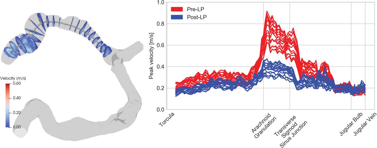
The peak velocity at several locations along the ipsilateral sinuses was obtained using in-house software, schematically described in Fig 1. Postprocessing begins with contouring the transverse and sigmoid sinuses on the symptomatic side from the CE-MRV. This is followed by a phase-offset background correction to the 4Dflow and registering the CE-MRV contour to both the pre- and post-LP 4Dflow. Then, the centerline of the contoured lumen is extracted, and cross-sectional cut planes are generated at 1-mm intervals along the flow lumen. Last, peak velocities are extracted from each of the cut planes along the centerline across time. The peak velocity of >0.5 m/s visually matched the appearance of a flow jet, so this velocity was used as a threshold value to determine the presence of jets.

Fig 1.
  • Download figure
  • Open in new tab
  • Download powerpoint
Fig 1.

Left, MR venogram (gray) of the bilateral transverse and sigmoid sinuses showing the centerline and transverse cut planes. Right, The peak velocity along the centerline is plotted for different time points in the cardiac cycle before (red) and after (blue) LP.

Flow rates were quantified using Segment (http://segment.heiberg.se)22 to evaluate the 2Dflow data acquired through the sigmoid sinus.

Statistical Analysis

Linear regression was performed to investigate the relation between the opening pressure and the change in peak velocity caused by the CSF drainage. To investigate potential confounding characteristics, we used ANOVA to test whether linear regression models with 2 variables were better than a model using the opening pressure alone. The additional variables investigated were flow rate, stenosis severity, change in CSF pressure, and the amount of CSF drained.

Statistical analysis was performed on both the group as a whole but also on the subgroup of patients with ipsilateral flow jets, which depict the impact for PT with suspected flow jet etiology.

Paired t tests were used to detect changes in intracranial pressure, flow rate, and velocities before and after lumbar puncture. The Pearson product momentum correlation was used to determine whether a reduction in peak velocity correlated with the reduction in intracranial pressure, and it was also used to determine whether the reduction in peak velocity correlated with the reduction in PT sound intensity.

Results

Ten patients were included in the study. Seven of the 10 patients did not meet the diagnostic criteria for IIH8; 2 were PT asymptomatic at the time of the pre-LP query; an ipsilateral jet with peak velocity of >0.5 m/s was identified in 8 of the 10 patients; and 1 patient underwent placement of a lumbar drain preventing recording of the closing pressure. A summary of the patient information is provided in Table 2.

View this table:
  • View inline
  • View popup
Table 2:

Summary of patientsa

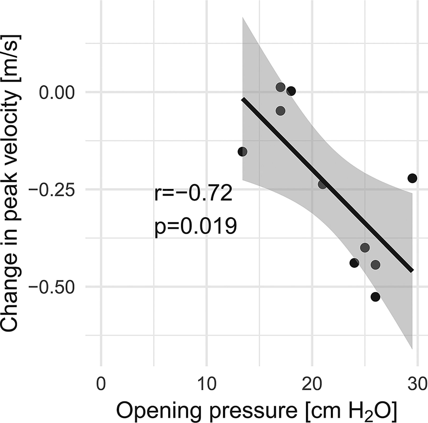
There was a correlation between the change in ipsilateral peak velocity and the opening pressure (r = −0.72, P = .019 for all patients, Fig 2, and r = −0.80, P = .030 for patients without IIH). No confounding characteristics were found by adding a second variable to the linear regression in addition to the opening pressure: flow rate (P = .208), stenosis severity (P = .375), change in CSF pressure (P = .543), and the amount of CSF drained (P = .529).

Fig 2.
  • Download figure
  • Open in new tab
  • Download powerpoint
Fig 2.

Lumbar puncture–induced change in ipsilateral peak velocity in relation to opening pressure.

No correlation was found between opening pressure and the change in the ipsilateral flow rate (r = 0.21, P = .564). There was also a correlation between the change in the PT rating and the reduction in peak velocity (r = 0.67, P = .035). The severity of the stenosis was not found to correlate with peak velocity (r = 0.50, P = .146) or a change in peak velocity (r = −0.40, P = .258).

Patients with an Ipsilateral Jet

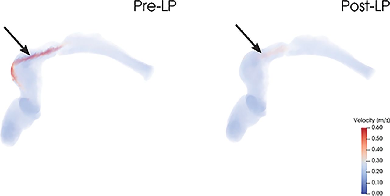
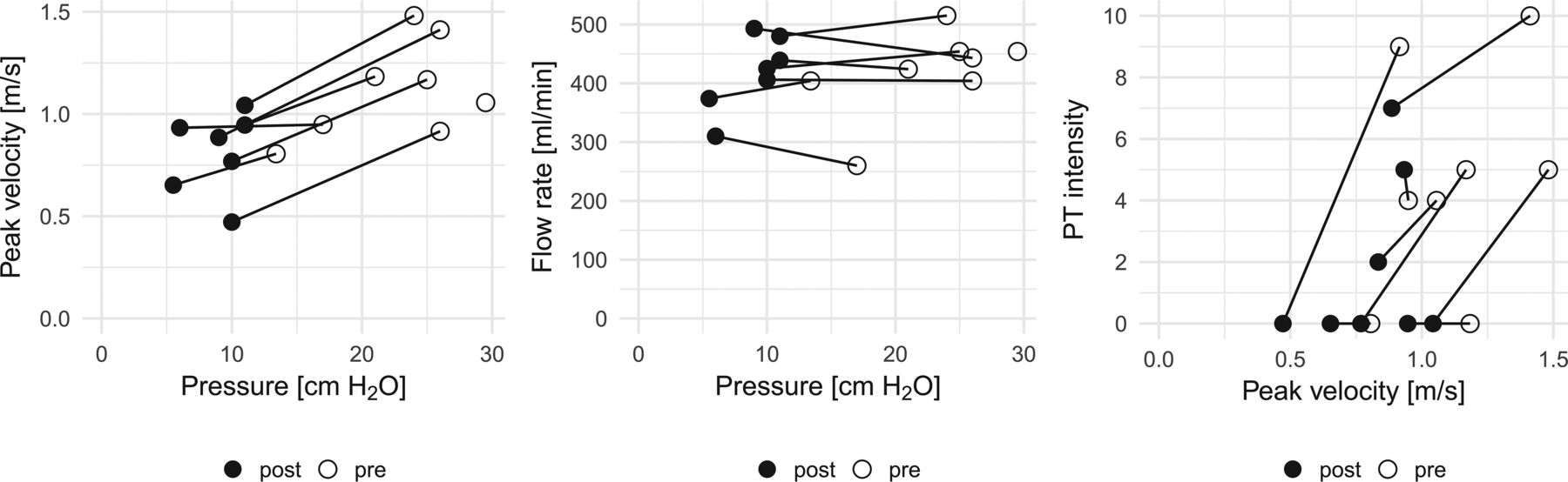
Eight of 10 patients had an ipsilateral flow jet, as illustrated in Fig 3. Two of these were asymptomatic at the time of the pre-LP query. The lumbar puncture reduced CSF pressure by a mean of 12.8 ± 3.4 cm H2O (P < .001) without significant changes in the ipsilateral bulk flow rate (P = .479). Nevertheless, the peak velocity was reduced by a mean of 0.30 ± 0.18 m/s (P = .002). The reduction in peak velocity correlated with the reduction in pressure (r = 0.82, P = .024) and with the reduction in subjectively scored PT intensity (r = 0.78, P = .023). The hemodynamic relations for these patients are illustrated in Fig 4.

Fig 3.
  • Download figure
  • Open in new tab
  • Download powerpoint
Fig 3.

The left side shows an ipsilateral flow jet pre-LP that is weakened post-LP without a concomitant change in the flow rate (374–381 mL/min). The rating of PT intensity for this patient was reduced from 9 to 0.

Fig 4.
  • Download figure
  • Open in new tab
  • Download powerpoint
Fig 4.

Lumbar puncture–induced hemodynamic changes for patients with ipsilateral jets. Note that reduced pressure lowered the ipsilateral peak velocity (left panel) without a concomitant reduction in flow rate (middle panel). Furthermore, PT intensity also decreased as the peak velocity was reduced.

The 6 patients experiencing pre-LP PT symptoms reported a mean PT intensity reduction of 3.8 ± 3.4.

Patients without an Ipsilateral Jet

Two patients did not have an ipsilateral flow jet. The first of these patients had a contralateral jet of reduced peak velocity after lumbar puncture (0.61 m/s from 0.67 m/s pre-LP), matching the mild improvement of the PT intensity (4 to 3). The second patient had high velocities in the external carotid artery, which subsided after LP, qualitatively matching the reduction in PT intensity (decreasing from 3 to 0).

Pre- and Post-LP MRV

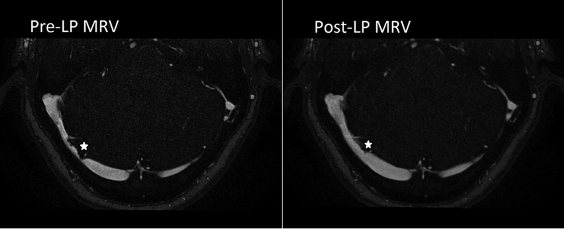
The patient imaged using ferumoxytol contrast underwent both pre- and post-LP CE-MR venography studies, demonstrating near-complete resolution of transverse sinus stenosis at a lower CSF pressure (10 cm H2O from 25 cm H2O before LP) (Fig 5).

Fig 5.
  • Download figure
  • Open in new tab
  • Download powerpoint
Fig 5.

LP-induced change in lumen geometry. The pre-LP MRV lumen is shown on the left side (opening pressure of 25 cm H2O), whereas the post-LP lumen is seen in the right side (closing pressure of 10 cm H2O). Note the improved transverse sinus stenosis at a prominent arachnoid granulation in the right transverse sinus (star).

Discussion

The aim of this study was to investigate changes in venous sinus flow features and PT intensity in response to lumbar puncture. The key findings in this study were the following: 1) Flow jets were seen in most cases in this study; 2) lowering the CSF pressure decreased the peak velocity of the flow jet in a venous sinus stenosis, without a concomitant reduction in the bulk flow rate; and 3) the reduction of peak velocity correlated with a reduced PT sound intensity. We thus hypothesize that the flow jet, and not the sinus bulk flow rate, is related to PT in patients with a transverse sinus stenosis.

The suspected cause of the decrease in peak jet velocity post-LP is an alleviation of transverse sinus stenosis as suggested by prior case reports.23⇓⇓–26 We were able to directly demonstrate this with MR venography in 1 patient using a blood pool contrast agent, and we believe this study is the first to use MR venography to show an immediate resolution in venous sinus stenosis after LP.

The direct relationship between peak jet velocity and CSF pressure was maintained for patients in this series who met the Dandy criteria for IIH (CSF opening pressure of >25 cm H2O) and for patients not meeting the Dandy criteria. Of our subjects who had improvement in their PT after removal of CSF, 5 of 7 had an opening pressure of ≥20 cm H2O, a threshold for abnormal elevation proposed by Corbett and Mehta27 for patients with a normal body mass index. Of note, venous sinus stenosis was observed in all patients in this series meeting or exceeding the 20-cm H2O threshold.

In a prospective trial, Boddu et al11 showed that PT can be alleviated in patients with IIH with venous sinus stent placement, and our results suggest that broadening this therapy to patients with PT who do not have IIH may warrant consideration, at least for patients with a transverse sinus stenosis whose symptoms improve with LP reduction of CSF pressure.28

While this study suggests the flow jet to be critical in the development of sound, the mechanistic link between a flow jet and PT symptoms remains opaque. We hypothesize that the transition into turbulent flow that can occur during the deceleration of these jets immediately “downstream” from the stenosis is a probable cause of the sound that causes PT.29 However, other anatomic variables may also play a role, such as sigmoid sinus diverticula, which can sometimes be seen in patients with transverse sinus stenosis.

At first glance, a finding that a reduction in symptoms following LP is not associated with a reduction in flow appears somewhat counterintuitive. Although not statistically significant, our data rather indicate that the flow rate might be slightly increased. On closer consideration however, it is known and expected that a vascular stenosis will, at first, have little effect on volume flow rates while peak velocities in the jet rise (and this feature forms the basis of Doppler sonography evaluation of stenoses of the extracranial carotid arteries). In carotid bruits, it is the disordered flow caused by the jet that is the source of sound, and that would be consistent with our observations reported here for PT. We also note that the reduced intracranial pressure seems to result in improvement in a very focal area of stenosis, and not the entirety of the sinus. In fact, most of the stenoses in this cohort were caused by compression from an arachnoid granulation as opposed to a smooth extrinsic stenosis. This discrepancy may help to explain the disparity between our results and prior work.

We hypothesized that the reduced peak velocities, without concomitant reduction in flow rates, primarily are the result of a reduction in the degree of stenosis. The stenoses caused by intracranial hypertension of >20 cm H2O are alleviated by an LP-induced reduction in intracranial pressure resulting in a concomitant reduction in the jet velocities. This hypothesis is supported by the pre- and post-LP MRA seen in Fig 5, which clearly shows widening of the transverse sinus stenosis after the LP.

Conclusions

The objective of this study was to evaluate changes in venous blood flow caused by lowered intracranial pressure in patients with PT after LP to help identify the cause of PT. Our results show an LP-induced reduction in venous peak velocity without a concomitant reduction in flow rate. We hypothesize that the reduction in jet velocity is caused by the alleviation of the transverse stenosis following LP. Our results further show a correlation between peak velocity and PT intensity, suggesting that flow jets are critical in the development of sound.

Acknowledgments

We thank Drs Wade Smith and Jonathan Horton for their helpful conversations in developing the concepts behind this work.

Footnotes

  • Disclosures: Henrik Haraldsson—RELATED: Grant: National Institutes of Health.* David A. Saloner—RELATED: Grant: National Institutes of Health.* Matthew R. Amans—RELATED: Grant: National Institutes of Health, Comments: R21DC016087*; UNRELATED: Consultancy: Covidien, Stryker Neurovascular, Comments: minor financial compensation for consultant work. Alexandra G. Wright—RELATED: Grant: National Institutes of Health, R21DC016087.* *Money paid to the institution.

  • This work was supported by National Institutes of Health, R21DC016087 (M.R.A./D.A.S.).

  • Paper previously presented, in part, at: Annual Meeting of the Society for Magnetic Resonance Angiography, August 28–31, 2018; Glasgow, Scotland; and American Society of Neuroradiology Annual Meeting and the Foundation of the ASNR Symposium, June 2–7, 2018; Vancouver, British Columbia, Canada.

Indicates open access to non-subscribers at www.ajnr.org

REFERENCES

  1. 1.↵
    1. Krishnan A,
    2. Mattox DE,
    3. Fountain AJ, et al
    . CT arteriography and venography in pulsatile tinnitus: preliminary results. AJNR Am J Neuroradiol 2006;27:1635–38 pmid:16971601
    Abstract/FREE Full Text
  2. 2.↵
    1. Liyanage SH,
    2. Singh A,
    3. Savundra P, et al
    . Pulsatile tinnitus. J Laryngol Otol 2006;120:93–97 doi:10.1017/S0022215105001714 pmid:16359136
    CrossRefPubMed
  3. 3.↵
    1. Madani G,
    2. Connor SE
    . Imaging in pulsatile tinnitus. Clin Radiol 2009;64:319–28 doi:10.1016/j.crad.2008.08.014 pmid:19185662
    CrossRefPubMed
  4. 4.↵
    1. Harvey RS,
    2. Hertzano R,
    3. Kelman SE, et al
    . Pulse-synchronous tinnitus and sigmoid sinus wall anomalies: descriptive epidemiology and the idiopathic intracranial hypertension patient population. Otol Neurotol 2014;35:7–15 doi:10.1097/MAO.0b013e3182a4756c pmid:24270723
    CrossRefPubMed
  5. 5.↵
    1. Cognard C,
    2. Gobin YP,
    3. Pierot L, et al
    . Cerebral dural arteriovenous fistulas: clinical and angiographic correlation with a revised classification of venous drainage. Radiology 1995;194:671–80 doi:10.1148/radiology.194.3.7862961 pmid:7862961
    CrossRefPubMed
  6. 6.↵
    1. Chan CF,
    2. Gatehouse PD,
    3. Hughes R, et al
    . Novel technique used to detect swallowing in volume-selective turbo spin-echo (TSE) for carotid artery wall imaging. J Magn Reson Imaging 2009;29:211–16 doi:10.1002/jmri.21607 pmid:19097078
    CrossRefPubMed
  7. 7.↵
    1. Olin JW,
    2. Froehlich J,
    3. Gu X, et al
    . The United States Registry for Fibromuscular Dysplasia: results in the first 447 patients. Circulation 2012;125:3182–90 doi:10.1161/CIRCULATIONAHA.112.091223 pmid:22615343
    Abstract/FREE Full Text
  8. 8.↵
    1. Morris PP,
    2. Black DF,
    3. Port J, et al
    . Transverse sinus stenosis is the most sensitive MR imaging correlate of idiopathic intracranial hypertension. AJNR Am J Neuroradiol 2017;38:471–77 doi:10.3174/ajnr.A5055 pmid:28104635
    Abstract/FREE Full Text
  9. 9.↵
    1. Morris PP,
    2. Lachman N,
    3. Black DF, et al
    . Increased curvature of the tentorium cerebelli in idiopathic intracranial hypertension. AJNR Am J Neuroradiol 2017;38:1789–93 doi:10.3174/ajnr.A5289 pmid:28663268
    Abstract/FREE Full Text
  10. 10.↵
    1. Farb RI,
    2. Vanek I,
    3. Scott JN, et al
    . Idiopathic intracranial hypertension: the prevalence and morphology of sinovenous stenosis. Neurology 2003;60:1418–24 doi:10.1212/01.WNL.0000066683.34093.E2 pmid:12743224
    Abstract/FREE Full Text
  11. 11.↵
    1. Boddu S,
    2. Dinkin M,
    3. Suurna M, et al
    . Resolution of pulsatile tinnitus after venous sinus stenting in patients with idiopathic intracranial hypertension. PLoS One 2016;11:e0164466 doi:10.1371/journal.pone.0164466 pmid:27768690
    CrossRefPubMed
  12. 12.↵
    1. Asif H,
    2. Craven CL,
    3. Siddiqui AH, et al
    . Idiopathic intracranial hypertension: 120-day clinical, radiological, and manometric outcomes after stent insertion into the dural venous sinus. J Neurosurg 2018;129:723–31 doi:10.3171/2017.4.JNS162871 pmid:28984521
    CrossRefPubMed
  13. 13.↵
    1. Dinkin MJ,
    2. Patsalides A
    . Venous sinus stenting in idiopathic intracranial hypertension: results of a prospective trial. J Neuroophthalmol 2017;37:113–21 doi:10.1097/WNO.0000000000000426 pmid:27556959
    CrossRefPubMed
  14. 14.↵
    1. Liu KC,
    2. Starke RM,
    3. Durst CR, et al
    . Venous sinus stenting for reduction of intracranial pressure in IIH: a prospective pilot study. J Neurosurg 2017;127:1126–33 doi:10.3171/2016.8.JNS16879 pmid:28009240
    CrossRefPubMed
  15. 15.↵
    1. Kao E,
    2. Kefayati S,
    3. Amans MR, et al
    . Flow patterns in the jugular veins of pulsatile tinnitus patients. J Biomech 2017;52:61–67 doi:10.1016/j.jbiomech.2016.12.008 pmid:28057349
    CrossRefPubMed
  16. 16.↵
    1. Kefayati S,
    2. Amans M,
    3. Faraji F, et al
    . The manifestation of vortical and secondary flow in the cerebral venous outflow tract: an in vivo MR velocimetry study. J Biomech 2017;50:180–87 doi:10.1016/j.jbiomech.2016.11.041 pmid:27894675
    CrossRefPubMed
  17. 17.↵
    1. Acevedo-Bolton G,
    2. Amans MR,
    3. Kefayati S, et al
    . Four dimensional magnetic resonance velocimetry for complex flow in the jugular vein. Quant Imaging Med Surg 2015;5:635–37 doi:10.3978/j.issn.2223-4292.2015.04.09 pmid:26435930
    CrossRefPubMed
  18. 18.↵
    1. Smith JL
    . Whence pseudotumor cerebri? J Clin Neuroophthalmol 1985;5:55–56 pmid:3156890
    PubMed
  19. 19.↵
    1. Ittrich H,
    2. Peldschus K,
    3. Raabe N, et al
    . Superparamagnetic iron oxide nanoparticles in biomedicine: applications and developments in diagnostics and therapy. Rofo 2013;185:1149–66 doi:10.1055/s-0033-1335438 pmid:24008761
    CrossRefPubMed
  20. 20.↵
    1. Weinstein JS,
    2. Varallyay CG,
    3. Dosa E, et al
    . Superparamagnetic iron oxide nanoparticles: diagnostic magnetic resonance imaging and potential therapeutic applications in neurooncology and central nervous system inflammatory pathologies: a review. J Cereb Blood Flow Metab 2010;30:15–35 doi:10.1038/jcbfm.2009.192 pmid:19756021
    CrossRefPubMed
  21. 21.↵
    1. Hope MD,
    2. Hope TA,
    3. Zhu C, et al
    . Vascular imaging with ferumoxytol as a contrast agent. AJR Am J Roentgenol 2015;205:W366–73 doi:10.2214/AJR.15.14534 pmid:26102308
    CrossRefPubMed
  22. 22.↵
    1. Heiberg E,
    2. Sjögren J,
    3. Ugander M, et al
    . Design and validation of Segment: freely available software for cardiovascular image analysis. BMC Med Imaging 2010;10:1 doi:10.1186/1471-2342-10-1 pmid:20064248
    CrossRefPubMed
  23. 23.↵
    1. Scoffings DJ,
    2. Pickard JD,
    3. Higgins JN
    . Resolution of transverse sinus stenoses immediately after CSF withdrawal in idiopathic intracranial hypertension. J Neurol Neurosurg Psychiatry 2007;78:911–12 doi:10.1136/jnnp.2006.111765 pmid:17635988
    FREE Full Text
  24. 24.↵
    1. King JO,
    2. Mitchell PJ,
    3. Thomson KR, et al
    . Manometry combined with cervical puncture in idiopathic intracranial hypertension. Neurology 2002;58:26–30 doi:10.1212/WNL.58.1.26 pmid:11781401
    Abstract/FREE Full Text
  25. 25.↵
    1. Baryshnik DB,
    2. Farb RI
    . Changes in the appearance of venous sinuses after treatment of disordered intracranial pressure. Neurology 2004;62:1445–46 doi:10.1212/01.WNL.0000120750.40453.64 pmid:15111701
    FREE Full Text
  26. 26.↵
    1. Onder H,
    2. Gocmen R,
    3. Gursoy-Ozdemir Y
    . Reversible transverse sinus collapse in a patient with idiopathic intracranial hypertension. BMJ Case Rep 2015;2015 doi:10.1136/bcr-2014-011606 pmid:25979956
    Abstract/FREE Full Text
  27. 27.↵
    1. Corbett JJ,
    2. Mehta MP
    . Cerebrospinal fluid pressure in normal obese subjects and patients with pseudotumor cerebri. Neurology 1983;33:1386–88 doi:10.1212/WNL.33.10.1386 pmid:6684240
    Abstract/FREE Full Text
  28. 28.↵
    1. Lenck S,
    2. Labeyrie MA,
    3. Vallee F, et al
    . Stent placement for disabling pulsatile tinnitus caused by a lateral sinus stenosis: a retrospective study. Oper Neurosurg (Hagerstown) 2017;13:560–65 doi:10.1093/ons/opx026 pmid:28922875
    CrossRefPubMed
  29. 29.↵
    1. Baomin L,
    2. Yongbing S,
    3. Xiangyu C
    . Angioplasty and stenting for intractable pulsatile tinnitus caused by dural venous sinus stenosis: a case series report. Otol Neurotol 2014;35:366–70 doi:10.1097/MAO.0b013e3182990d52 pmid:24080976
    CrossRefPubMed
  • Received August 1, 2018.
  • Accepted after revision March 18, 2019.
  • © 2019 by American Journal of Neuroradiology
View Abstract
PreviousNext
Back to top

In this issue

American Journal of Neuroradiology: 40 (5)
American Journal of Neuroradiology
Vol. 40, Issue 5
1 May 2019
  • Table of Contents
  • Index by author
  • Complete Issue (PDF)
Advertisement
Print
Download PDF
Email Article

Thank you for your interest in spreading the word on American Journal of Neuroradiology.

NOTE: We only request your email address so that the person you are recommending the page to knows that you wanted them to see it, and that it is not junk mail. We do not capture any email address.

Enter multiple addresses on separate lines or separate them with commas.
Reduced Jet Velocity in Venous Flow after CSF Drainage: Assessing Hemodynamic Causes of Pulsatile Tinnitus
(Your Name) has sent you a message from American Journal of Neuroradiology
(Your Name) thought you would like to see the American Journal of Neuroradiology web site.
CAPTCHA
This question is for testing whether or not you are a human visitor and to prevent automated spam submissions.
Cite this article
H. Haraldsson, J.R. Leach, E.I. Kao, A.G. Wright, S.G. Ammanuel, R.S. Khangura, M.K. Ballweber, C.T. Chin, V.N. Shah, K. Meisel, D.A. Saloner, M.R. Amans
Reduced Jet Velocity in Venous Flow after CSF Drainage: Assessing Hemodynamic Causes of Pulsatile Tinnitus
American Journal of Neuroradiology May 2019, 40 (5) 849-854; DOI: 10.3174/ajnr.A6043

Citation Manager Formats

  • BibTeX
  • Bookends
  • EasyBib
  • EndNote (tagged)
  • EndNote 8 (xml)
  • Medlars
  • Mendeley
  • Papers
  • RefWorks Tagged
  • Ref Manager
  • RIS
  • Zotero
0 Responses
Respond to this article
Share
Bookmark this article
Reduced Jet Velocity in Venous Flow after CSF Drainage: Assessing Hemodynamic Causes of Pulsatile Tinnitus
H. Haraldsson, J.R. Leach, E.I. Kao, A.G. Wright, S.G. Ammanuel, R.S. Khangura, M.K. Ballweber, C.T. Chin, V.N. Shah, K. Meisel, D.A. Saloner, M.R. Amans
American Journal of Neuroradiology May 2019, 40 (5) 849-854; DOI: 10.3174/ajnr.A6043
del.icio.us logo Twitter logo Facebook logo Mendeley logo
  • Tweet Widget
  • Facebook Like
  • Google Plus One
Purchase

Jump to section

  • Article
    • Abstract
    • ABBREVIATIONS:
    • Materials and Methods
    • Results
    • Discussion
    • Conclusions
    • Acknowledgments
    • Footnotes
    • REFERENCES
  • Figures & Data
  • Info & Metrics
  • Responses
  • References
  • PDF

Related Articles

  • No related articles found.
  • PubMed
  • Google Scholar

Cited By...

  • Pulsatile Tinnitus Due to Stenosis of the Marginal Sinus: Diagnosis and Endovascular Treatment
  • Crossref (21)
  • Google Scholar

This article has been cited by the following articles in journals that are participating in Crossref Cited-by Linking.

  • Diagnostic Approach to Pulsatile Tinnitus
    Kazim H. Narsinh, Ferdinand Hui, David Saloner, Adelyn Tu-Chan, Jeffrey Sharon, Andreas M. Rauschecker, Fatima Safoora, Vinil Shah, Karl Meisel, Matthew R. Amans
    JAMA Otolaryngology–Head & Neck Surgery 2022 148 5
  • A proposed framework for cerebral venous congestion
    Anirudh Arun, Matthew R Amans, Nicholas Higgins, Waleed Brinjikji, Mithun Sattur, Sudhakar R Satti, Peter Nakaji, Mark Luciano, Thierry AGM Huisman, Abhay Moghekar, Vitor M Pereira, Ran Meng, Kyle Fargen, Ferdinand K Hui
    The Neuroradiology Journal 2022 35 1
  • Why does unilateral pulsatile tinnitus occur in patients with idiopathic intracranial hypertension?
    Pengfei Zhao, Chenyu Jiang, Han Lv, Tong Zhao, Shusheng Gong, Zhenchang Wang
    Neuroradiology 2021 63 2
  • Correlation Between Trans-Stenotic Blood Flow Velocity Differences and the Cerebral Venous Pressure Gradient in Transverse Sinus Stenosis: A Prospective 4-Dimensional Flow Magnetic Resonance Imaging Study
    Heyu Ding, Pengfei Zhao, Han Lv, Xiaoshuai Li, Xiaoyu Qiu, Rong Zeng, Guopeng Wang, Zhenghan Yang, Shusheng Gong, Long Jin, Zhenchang Wang
    Neurosurgery 2021 89 4
  • From2Dto4DPhase‐ContrastMRIin the Neurovascular System: Will It Be a Quantum Jump or a Fancy Decoration?
    Sung Won Youn, Jongmin Lee
    Journal of Magnetic Resonance Imaging 2022 55 2
  • Evidence of air‐conduction transmission pathway and strategized transtemporal operative techniques for venous pulsatile tinnitus: Combining water occlusion test and operative sensing applications
    Yue‐Lin Hsieh, Xiaobing Xu, Yongzhen Wu, Wuqing Wang
    Laryngoscope Investigative Otolaryngology 2021 6 6
  • Pulsatile Tinnitus Due to Stenosis of the Marginal Sinus: Diagnosis and Endovascular Treatment
    J. Cortese, M. Eliezer, A. Guédon, E. Houdart
    American Journal of Neuroradiology 2021 42 12
  • Emergence of Venous Stenosis as the Dominant Cause of Pulsatile Tinnitus
    Eytan Raz, Erez Nossek, Daniel Jethanamest, Vinayak Narayan, Aryan Ali, Vera Sharashidze, Tibor Becske, Peter K. Nelson, Maksim Shapiro
    Stroke: Vascular and Interventional Neurology 2022 2 4
  • Intracranial pressure, lateral sinus patency, and jugular ultrasound hemodynamics in patients with venous pulsatile tinnitus
    Xiuli Gao, Yue-Lin Hsieh, Shenjiang Wang, Suming Shi, Wuqing Wang
    Frontiers in Neurology 2022 13
  • Brain herniation (encephalocele) into arachnoid granulations: prevalence and association with pulsatile tinnitus and idiopathic intracranial hypertension
    Eric R. Smith, M. Travis Caton, Javier E. Villanueva-Meyer, Justin Remer, Laura B. Eisenmenger, Amanda Baker, Vinil N. Shah, Adelyn Tu-Chan, Karl Meisel, Matthew R. Amans
    Neuroradiology 2022 64 9

More in this TOC Section

  • WHO Classification Update: Nasal&Skull Base Tumors
  • Peritumoral Signal in Vestibular Schwannomas
  • Chondrosarcoma vs Synovial Chondromatosis: Imaging
Show more HEAD & NECK

Similar Articles

Advertisement

Indexed Content

  • Current Issue
  • Accepted Manuscripts
  • Article Preview
  • Past Issues
  • Editorials
  • Editors Choice
  • Fellow Journal Club
  • Letters to the Editor

Cases

  • Case Collection
  • Archive - Case of the Week
  • Archive - Case of the Month
  • Archive - Classic Case

Special Collections

  • Special Collections

Resources

  • News and Updates
  • Turn around Times
  • Submit a Manuscript
  • Author Policies
  • Manuscript Submission Guidelines
  • Evidence-Based Medicine Level Guide
  • Publishing Checklists
  • Graphical Abstract Preparation
  • Imaging Protocol Submission
  • Submit a Case
  • Become a Reviewer/Academy of Reviewers
  • Get Peer Review Credit from Publons

Multimedia

  • AJNR Podcast
  • AJNR SCANtastic
  • Video Articles

About Us

  • About AJNR
  • Editorial Board
  • Not an AJNR Subscriber? Join Now
  • Alerts
  • Feedback
  • Advertise with us
  • Librarian Resources
  • Permissions
  • Terms and Conditions

American Society of Neuroradiology

  • Not an ASNR Member? Join Now

© 2025 by the American Society of Neuroradiology All rights, including for text and data mining, AI training, and similar technologies, are reserved.
Print ISSN: 0195-6108 Online ISSN: 1936-959X

Powered by HighWire