
Article Figures & Data
Figures
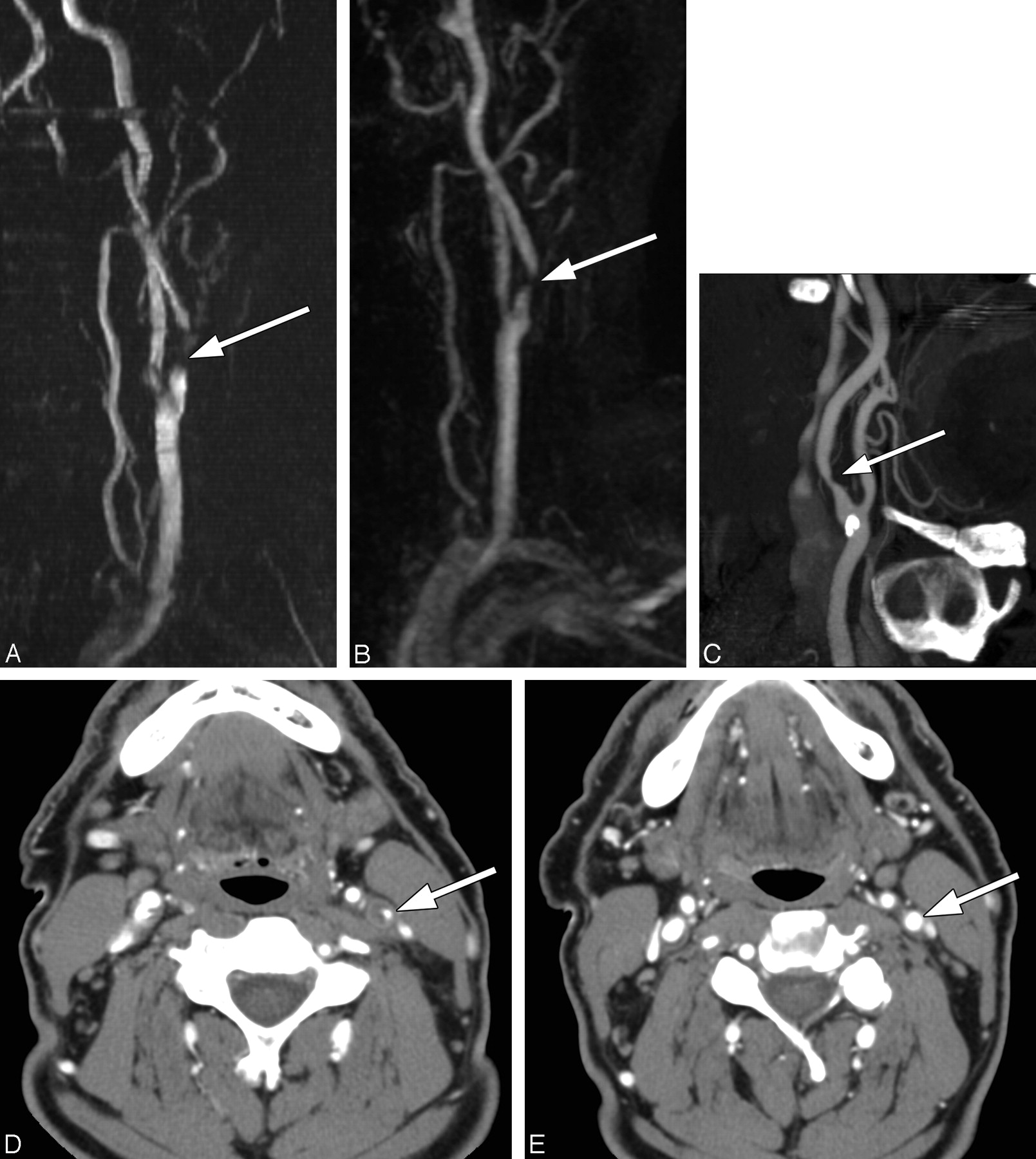
- Fig 1.
This ICA origin was rated by the observers to have severe (70%–95%) stenosis when imaged by all 3 techniques: 2D TOF MRA (A), CEMRA (B), and CTA (C–E). A and B, Signal-intensity drop-out is noted (arrow), but no distal narrowing or distal signal-intensity reduction is observed on MRA images. C, Curved reformatted CTA view of the left ICA demonstrating a severe stenosis (arrow). D, An axial image at the level of greatest narrowing (arrow). E, At the level of the NASCET reference diameter (arrow).
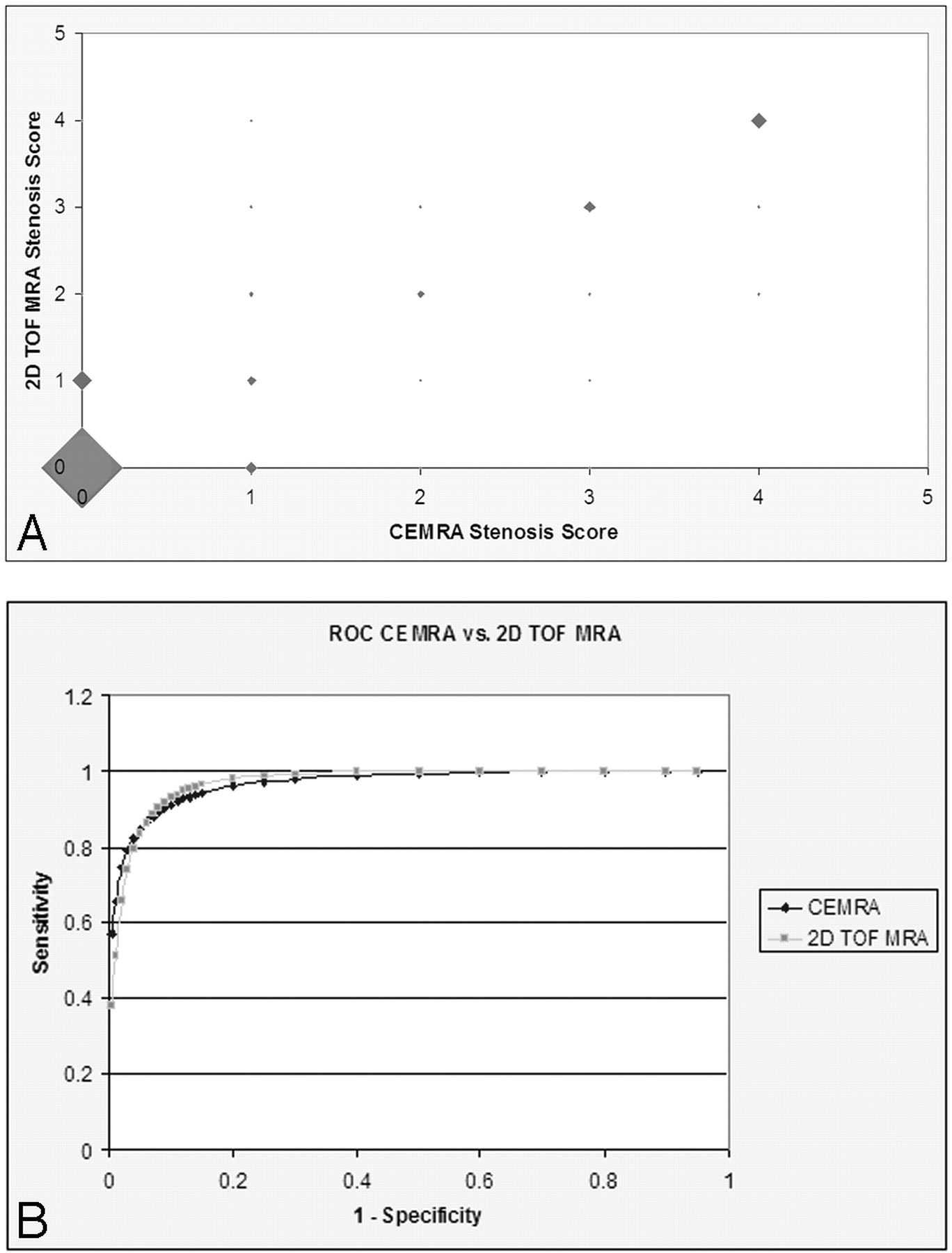
- Fig 2.
A scatterplot of stenosis scores on CE-MRA and 2D TOF MRA (A) and ROC curves for CE-MRA versus 2D TOF MRA (B). The size of the marker on the scatterplot represents the relative frequency of stenosis scores.
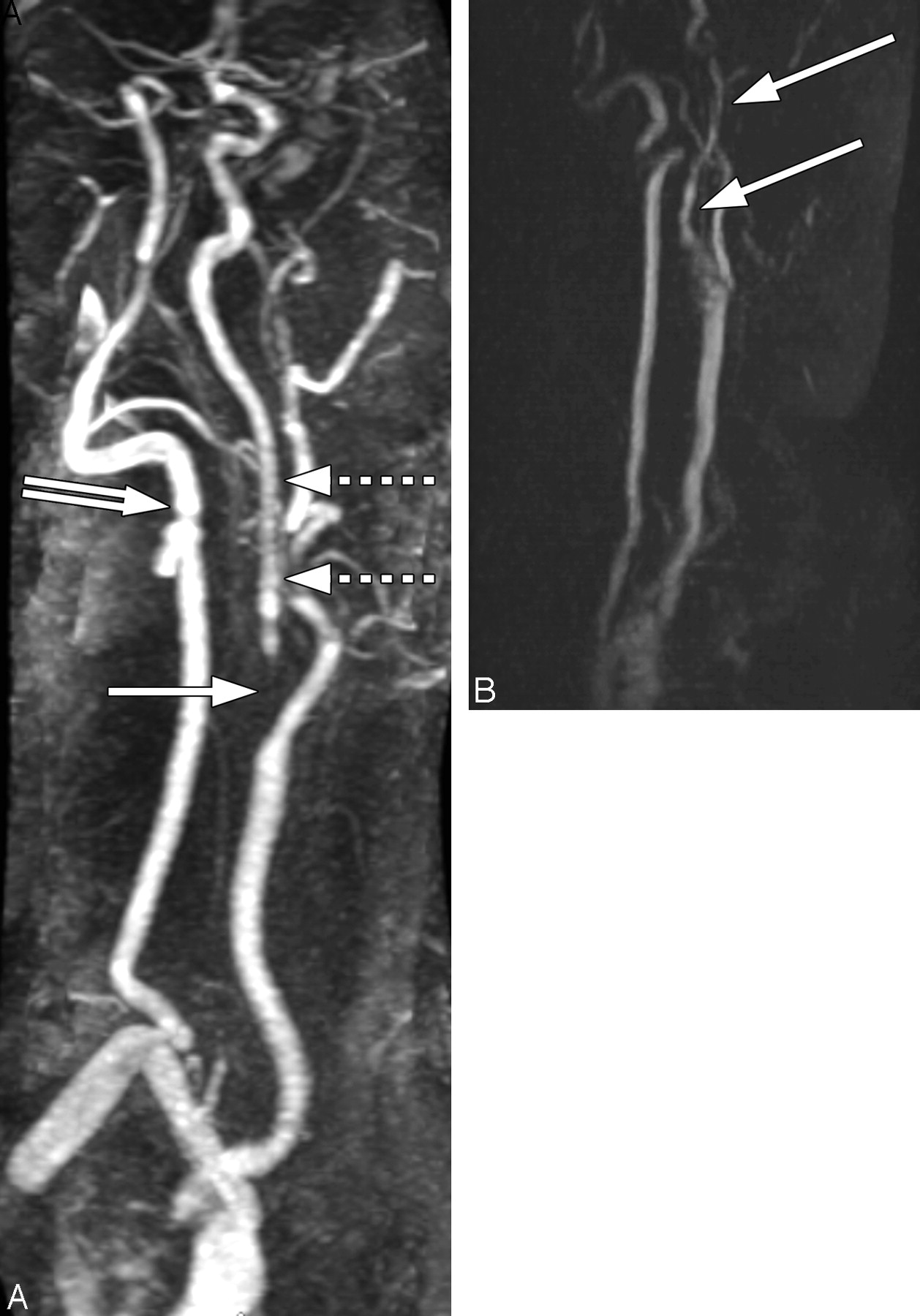
- Fig 3.
A, MRA of the neck with gadolinium demonstrating signal-intensity drop-out in the proximal left ICA (solid arrow), with decreased signal intensity and vessel narrowing (slim sign) in the distal ICA (dashed arrows), which is significantly smaller compared with the ipsilateral vertebral artery (double-tailed arrow). B, 2D TOF MRA image of the neck vasculature exemplifies distal-vessel narrowing/irregularity and distal-vessel signal-intensity reduction (white arrows).


Tables
- Table 1:
Comparison of CTA versus CE-MRA, CTA versus 2D TOF MRA, and CE-MRA versus 2D TOF MRA stenosis scores
Techniques Difference None By 1 By 2 By 3 By 4 CTA vs CE-MRA 265/354 (75%) 72/354 (20%) 13/354 (4%) 2/354 (0.5%) 2/354 (0.5%) CTA vs 2D TOF MRA 227/354 (64%) 104/354 (29%) 20/354 (6%) 0/354 (0%) 3/354 (1%) CE-MRA vs 2D TOF MRA 250/354 (71%) 85/354 (24%) 15/354 (4%) 3/354 (0.75%) 1/354 (0.25%) Note:—CTA indicates CT angiography; CE-MRA, contrast-enhanced MR angiography; TOF MRA, time-of-flight MR angiography.
- Table 2:
Sensitivity, specificity, accuracy, PPV, and NPV of CE-MRA and unenhanced 2D TOF MRA for detection of ≥70% ICA stenosis* based on CTA reference measurements
Technique Sensitivity Specificity Accuracy PPV NPV CE-MRA 83.6% (46/55) 95.7% (286/299) 93.8% (332/354) 78.0% (46/59) 96.9% (286/295) 2D TOF MRA 80.0% (44/55) 95.3% (285/299) 92.9% (329/354) 75.9% (44/58) 96.3% (285/296) Note:—PPV indicates positive predictive value; NPV, negative predictive value; ICA, internal carotid artery.
* Scores of 3 and 4.
- Table 3:
Sensitivity, specificity, accuracy, PPV, and NPV of signal drop-out, distal-vessel narrowing, and distal-vessel signal-intensity reduction for detection of ≥70% ICA stenosis* based on CTA reference measurements
Sensitivity Specificity Accuracy PPV NPV CE-MRA Signal drop-out 65.5% (36/55) 98.3% (294/299) 93.2% (330/354) 87.8% (36/41) 93.9% (294/313) Distal-vessel narrowing 41.8% (23/55) 99.3% (297/299) 90.4% (320/354) 92% (23/25) 90.3% (297/329) Distal-vessel signal reduction 41.8% (23/55) 99.3% (297/299) 90.4% (320/354) 92% (23/25) 90.3% (297/329) 2D TOF MRA Signal drop-out 50.9% (28/55) 98.7% (295/299) 91.2% (323/354) 87.5% (28/32) 91.6% (295/322) Distal-vessel narrowing 34.5% (19/55) 99% (296/299) 89% (315/354) 86.4% (19/22) 89.2% (296/332) Distal-vessel signal reduction 36.4% (20/55) 99% (296/299) 89.3% (316/354) 87% (20/23) 89.4% (296/331) * Scores of 3 and 4.
In this issue


Jump to section
Related Articles
Cited By...
- Intraplaque High-Intensity Signal on 3D Time-of-Flight MR Angiography Is Strongly Associated with Symptomatic Carotid Artery Stenosis
- Carotid CTA: Radiation Exposure and Image Quality with the Use of Attenuation-Based, Automated Kilovolt Selection
- Imaging challenges of carotid artery in-stent restenosis