Advertisement

Main menu

  • Home
  • Content
    • Current Issue
    • Accepted Manuscripts
    • Article Preview
    • Past Issue Archive
    • AJNR Case Collection
    • Case of the Week Archive
    • Classic Case Archive
    • Case of the Month Archive
  • Special Collections
    • Spinal CSF Leak Articles (Jan 2020-June 2024)
    • 2024 AJNR Journal Awards
    • Most Impactful AJNR Articles
  • Multimedia
    • AJNR Podcast
    • AJNR Scantastics
    • Video Articles
  • For Authors
    • Submit a Manuscript
    • Author Policies
    • Fast publishing of Accepted Manuscripts
    • Graphical Abstract Preparation
    • Manuscript Submission Guidelines
    • Imaging Protocol Submission
    • Submit a Case for the Case Collection
  • About Us
    • About AJNR
    • Editorial Board
  • More
    • Become a Reviewer/Academy of Reviewers
    • Subscribers
    • Permissions
    • Alerts
    • Feedback
    • Advertisers
    • ASNR Home
  • Other Publications
    • ajnr

User menu

  • Alerts
  • Log in

Search

  • Advanced search
American Journal of Neuroradiology

American Journal of Neuroradiology

ASHNR American Society of Functional Neuroradiology ASHNR American Society of Pediatric Neuroradiology ASSR
  • Alerts
  • Log in

Advanced Search

  • Home
  • Content
    • Current Issue
    • Accepted Manuscripts
    • Article Preview
    • Past Issue Archive
    • AJNR Case Collection
    • Case of the Week Archive
    • Classic Case Archive
    • Case of the Month Archive
  • Special Collections
    • Spinal CSF Leak Articles (Jan 2020-June 2024)
    • 2024 AJNR Journal Awards
    • Most Impactful AJNR Articles
  • Multimedia
    • AJNR Podcast
    • AJNR Scantastics
    • Video Articles
  • For Authors
    • Submit a Manuscript
    • Author Policies
    • Fast publishing of Accepted Manuscripts
    • Graphical Abstract Preparation
    • Manuscript Submission Guidelines
    • Imaging Protocol Submission
    • Submit a Case for the Case Collection
  • About Us
    • About AJNR
    • Editorial Board
  • More
    • Become a Reviewer/Academy of Reviewers
    • Subscribers
    • Permissions
    • Alerts
    • Feedback
    • Advertisers
    • ASNR Home
  • Follow AJNR on Twitter
  • Visit AJNR on Facebook
  • Follow AJNR on Instagram
  • Join AJNR on LinkedIn
  • RSS Feeds

Welcome to the new AJNR, Updated Hall of Fame, and more. Read the full announcements.


AJNR is seeking candidates for the position of Associate Section Editor, AJNR Case Collection. Read the full announcement.


Research ArticleInterventional

Image Quality of Low-Dose Cerebral Angiography and Effectiveness of Clinical Implementation on Diagnostic and Neurointerventional Procedures for Intracranial Aneurysms

J. Choi, B. Kim, Y. Choi, N.Y. Shin, J. Jang, H.S. Choi, S.L. Jung and K.J. Ahn
American Journal of Neuroradiology May 2019, 40 (5) 827-833; DOI: https://doi.org/10.3174/ajnr.A6029
J. Choi
aFrom the Department of Radiology (J.C.), St. Vincent's Hospital, College of Medicine, The Catholic University of Korea, Gyeonggi-do, Republic of Korea
  • Find this author on Google Scholar
  • Find this author on PubMed
  • Search for this author on this site
  • ORCID record for J. Choi
B. Kim
bDepartment of Radiology (B.K., Y.C., N.Y.S., J.J., H.S.C., S.L.J., K.J.A.), Seoul St. Mary's Hospital, College of Medicine, The Catholic University of Korea, Seoul, Republic of Korea.
  • Find this author on Google Scholar
  • Find this author on PubMed
  • Search for this author on this site
  • ORCID record for B. Kim
Y. Choi
bDepartment of Radiology (B.K., Y.C., N.Y.S., J.J., H.S.C., S.L.J., K.J.A.), Seoul St. Mary's Hospital, College of Medicine, The Catholic University of Korea, Seoul, Republic of Korea.
  • Find this author on Google Scholar
  • Find this author on PubMed
  • Search for this author on this site
  • ORCID record for Y. Choi
N.Y. Shin
bDepartment of Radiology (B.K., Y.C., N.Y.S., J.J., H.S.C., S.L.J., K.J.A.), Seoul St. Mary's Hospital, College of Medicine, The Catholic University of Korea, Seoul, Republic of Korea.
  • Find this author on Google Scholar
  • Find this author on PubMed
  • Search for this author on this site
  • ORCID record for N.Y. Shin
J. Jang
bDepartment of Radiology (B.K., Y.C., N.Y.S., J.J., H.S.C., S.L.J., K.J.A.), Seoul St. Mary's Hospital, College of Medicine, The Catholic University of Korea, Seoul, Republic of Korea.
  • Find this author on Google Scholar
  • Find this author on PubMed
  • Search for this author on this site
  • ORCID record for J. Jang
H.S. Choi
bDepartment of Radiology (B.K., Y.C., N.Y.S., J.J., H.S.C., S.L.J., K.J.A.), Seoul St. Mary's Hospital, College of Medicine, The Catholic University of Korea, Seoul, Republic of Korea.
  • Find this author on Google Scholar
  • Find this author on PubMed
  • Search for this author on this site
  • ORCID record for H.S. Choi
S.L. Jung
bDepartment of Radiology (B.K., Y.C., N.Y.S., J.J., H.S.C., S.L.J., K.J.A.), Seoul St. Mary's Hospital, College of Medicine, The Catholic University of Korea, Seoul, Republic of Korea.
  • Find this author on Google Scholar
  • Find this author on PubMed
  • Search for this author on this site
  • ORCID record for S.L. Jung
K.J. Ahn
bDepartment of Radiology (B.K., Y.C., N.Y.S., J.J., H.S.C., S.L.J., K.J.A.), Seoul St. Mary's Hospital, College of Medicine, The Catholic University of Korea, Seoul, Republic of Korea.
  • Find this author on Google Scholar
  • Find this author on PubMed
  • Search for this author on this site
  • ORCID record for K.J. Ahn
  • Article
  • Figures & Data
  • Info & Metrics
  • Responses
  • References
  • PDF
Loading

Abstract

BACKGROUND AND PURPOSE: Awareness of the potential for exposure to high doses of radiation from interventional radiologic procedures has increased. The purpose of this study was to evaluate image quality and dose reduction of low-dose cerebral angiography during diagnostic and therapeutic procedures for intracranial aneurysms.

MATERIALS AND METHODS: A retrospective review of 1137 prospectively collected patients between January 2012 and June 2014 was performed. Beginning in April 2013, a dose-reduction strategy was implemented. Subjective image-quality assessment of 506 standard and 540 low-dose cerebral angiography images was performed by 2 neuroradiologists using a 5-point scale and was tested using noninferiority statistics. Radiation dose-area product and air kerma of 1046 diagnostic and 317 therapeutic procedures for intracranial aneurysms were analyzed and compared between groups before (group 1) and after (group 2) clinical implementation of a dose-reduction strategy.

RESULTS: The image quality of the low-dose cerebral angiography was not inferior on the basis of results from the 2 readers. For diagnostic cerebral angiography, the mean dose-area product and air kerma were 140.8 Gy×cm2 and 1.0 Gy, respectively, in group 1 and 82.0 Gy×cm2 and 0.6 Gy in group 2 (P < .001, P < .001). For the neurointerventional procedure, the mean dose-area product and air kerma were 246.0 Gy×cm2 and 3.7 Gy, respectively, in group 1 and 169.8 Gy×cm2 and 3.3 Gy in group 2 (P < .001, P = .291).

CONCLUSIONS: With low-dose cerebral angiography, image quality was maintained, and implementation of dose-reduction strategies reduced radiation doses in patients undergoing diagnostic and neurointerventional procedures for intracranial aneurysms.

ABBREVIATIONS:

AK
air kerma
DAP
dose-area product

Although CT and MR angiography are performed frequently to diagnose cerebrovascular diseases, cerebral angiography is still the criterion standard for the evaluation of the vasculature in cerebrovascular disease, especially when therapeutic procedures are anticipated. The potential for high radiation doses from interventional radiologic procedures is well-known; thus, such procedures should only be performed with adequate justification.1,2 The adverse health effects of radiation exposure can be divided into 2 categories: deterministic effects and stochastic effects. Skin dose is related to deterministic effects such as skin erythema, necrosis, ulceration, cataract, and permanent epilation.1,3 Although direct measurement of the skin radiation dose using thermoluminescent dosimeters is the most accurate, it is not practical because it is a cumbersome procedure with limitations.4,5 Dose-area product (DAP) and air kerma (AK) are alternative indirect measurements of a patient dose and are the most convenient and currently most widely used parameters for monitoring it.4,6⇓⇓⇓–10

With increased awareness of the need for reduction of the radiation dose during cerebral angiography, many techniques and strategies have reduced unnecessary radiation doses while maintaining image quality needed to perform procedures.7⇓⇓⇓⇓–12 At Seoul St. Mary's Hospital, we have recorded radiation parameters for each procedure since 2009, and our early experience has shown that the proportion of the radiation dose in terms of DAP from angiographic acquisitions was 88.6% for diagnostic cerebral angiography and 49.5% for neurointerventional procedures.6 Thus, low-dose angiography provides good image quality and may be an effective way to reduce the radiation dose to patients undergoing diagnostic or neurointerventional procedures. Although there have been several reports showing the effectiveness of low-dose cerebral angiography, little work has been done to study the application and its effectiveness in a clinical setting with a large volume of patients.8,9,13⇓–15

Since April 2013, our clinic has implemented the following new dose-reduction strategy: 1) low-dose angiographic protocol (from 3.6 μGy/frame to 1.8 μGy/frame) for cerebral angiography, and 2) a roadmap image saved for evaluation of the femoral arterial puncture site, instead of femoral angiography. The purpose of this study was to evaluate image quality of low-dose cerebral angiography and the effectiveness of the clinical implementation of our dose-reduction strategies in diagnostic and neurointerventional procedures for intracranial aneurysms.

Materials and Methods

This study was approved by Seoul St. Mary's Hospital institutional review board. A waiver of the need for consent was obtained for this Health Insurance Portability and Accountability Act–compliant retrospective study. Retrospective review of the prospectively collected data base of 1137 patients (799 females, 338 males; median age, 56 years; range, 13–88 years) between January 2012 and June 2014 was performed. All procedures were performed by a team of 2 experienced neurointerventionalists. A clinical fellow with >300 previous cases performed the diagnostic angiographic procedures, while embolizations were performed by a radiologist with >20 years of experience in aneurysm embolization. Diagnostic procedures performed for follow-up after clipping of an aneurysm were excluded. There were, in total, 1046 diagnostic and 317 therapeutic procedures.

The angiographic system used was a biplane angiographic unit (Axiom Artis dBA; Siemens, Erlangen, Germany) with a flat panel detector: A and B planes (48 cm) with variable FOVs of 42 to 32–22 to 16 cm. The total filtration was 2.5 mm Al. The angiography unit had 3 pulsed fluoroscopy modes of 10, 15, and 30 P/s, of which 15 P/s was used most frequently with variable copper filtration of up to 0.9 mm. The system had a DAP meter, including a verification of the AK and DAP displayed values against an external dosimeter during the quality-assurance process at least once a year.

Since April 2013, our institution has applied the following dose-reduction strategy: 1) low-dose angiographic protocol (from 3.6 μGy/frame to 1.8 μGy/frame) for cerebral angiography, and 2) a roadmap image saved for evaluation of the femoral artery puncture site, instead of femoral angiography. Angiographic procedures were the same as those used in a previous protocol, including acquisition of internal carotid and vertebral arteriographies in anteroposterior and lateral views, and rotational angiography was performed using 5-second dual runs with 0.36 μGy/frame when an intracranial aneurysm was suspected on 2D cerebral angiography.6

Two experienced neuroradiologists with >15 years of experience evaluated the quality of the images, which had been arranged in a random order. They reviewed all DSA series, and they were blinded to the examination date to avoid knowing the dose category. They analyzed the image quality on a scale of 5, with the higher number indicating better image quality: 1 (nondiagnostic), 2 (poor: compromised large and small vessel visualization), 3 (average: diagnostic value for large vessel but compromised small vessel visualization), 4 (good: excellent large vessel and minimal compromise of small vessel visualization), 5 (very good: excellent large and small vessel visualization).

The parameters measured were DAP, AK, fluoroscopy exposure time, and the number of angiographic image acquisitions for all procedures. Image quality was also obtained for 1046 diagnostic procedures (506 procedures before the low-dose protocol and 540 procedures after the low-dose protocol).

A noninferiority test was used for the image quality of diagnostic procedures before and after dose reduction, and interrater reliability was tested using the Cronbach α. The difference in parameters before and after the dose-reduction protocol was tested with a Student t test. SPSS (IBM, Armonk, New York) was used for statistical analysis. Statistical significance was defined as P < .05.

Results

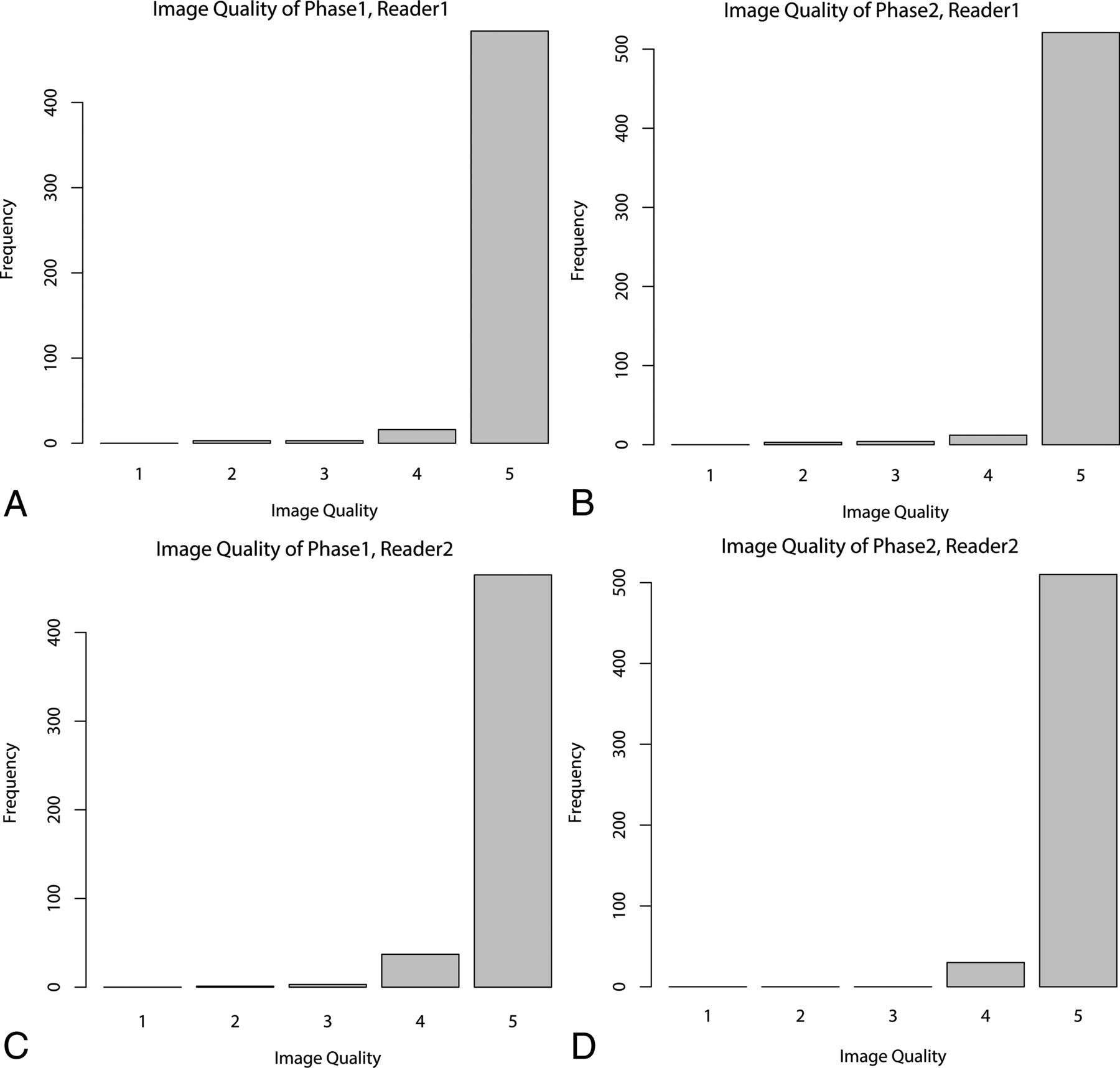
The subjective data for the quality of images before and after the low-dose protocol by 2 radiologists are presented in Table 1 and Fig 1. The image-quality score was well-correlated between the 2 observers (Cronbach α = 0.853). The diagnostic noninferiority of the angiography with 1.8 μGy/frame was demonstrated using the noninferiority test because the upper margin of the 1-sided 97.5% confidence interval did not cross the predefined noninferiority margin of −0.2. Figure 2 shows representative examples of the 2 cohorts.

View this table:
  • View inline
  • View popup
Table 1:

Mean image quality of angiography by 2 separate readers before and after the low-dose angiographic protocol

Fig 1.
  • Download figure
  • Open in new tab
  • Download powerpoint
Fig 1.

Image quality of angiography by 2 separate readers before (Phase 1) and after (Phase 2) the low-dose angiographic protocol.

Fig 2.
  • Download figure
  • Open in new tab
  • Download powerpoint
Fig 2.

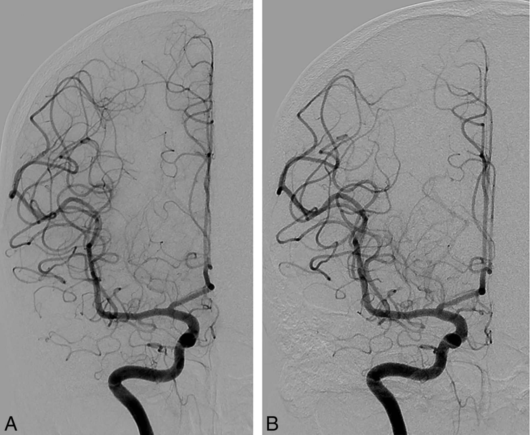
Representative examples of angiographic images before (A) and after (B) the low-dose angiographic protocol. These are cerebral angiograms of a patient who underwent diagnostic angiography to determine the cause of perimesencephalic subarachnoid hemorrhage.

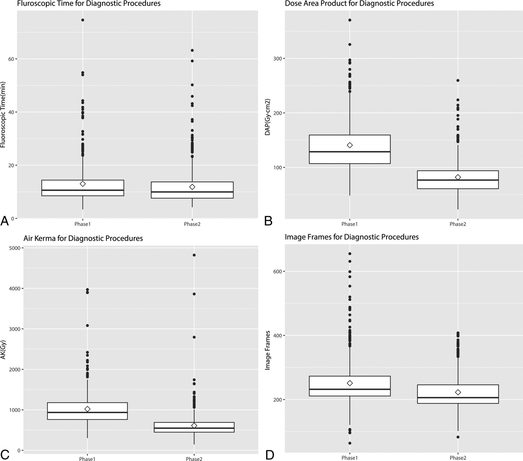
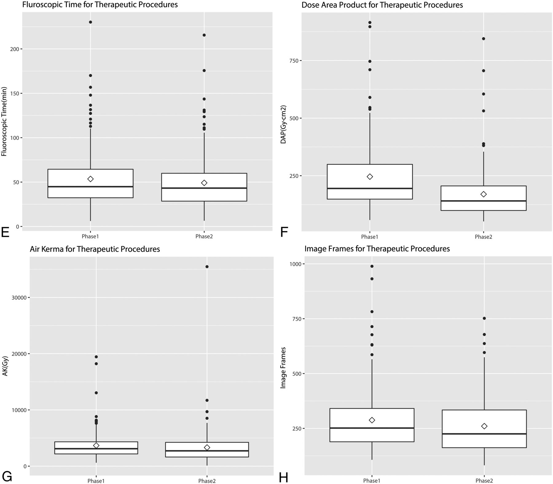
The quantitative data are shown in Table 2 and Fig 3. Before application of the low-dose angiographic protocol (Phase 1), mean fluoroscopic time, total mean DAP, total AK, and total angiographic image frames were 13.0 ± 7.9 minutes, 140.8 ± 48.1 Gy×cm2, 1.02 ± 0.42 Gy, and 251.2 ± 72 frames for diagnostic procedures, respectively, and 53.5 ± 34.1 minutes, 246.0 ± 148.3 Gy×cm2, 3.67 ± 2.66 Gy, and 287.6 ± 150 frames for therapeutic procedures. The third quartiles, which may be used as a dose reference level, were 14.4 and 64.5 minutes for fluoroscopy times, 160.8 and 301.4 Gy×cm2 for DAP, 1.18 and 4.42 Gy for AK, and 273 and 341 image frames in diagnostic and therapeutic procedures, respectively. After application of the low-dose angiographic protocol (Phase 2), mean fluoroscopic time, total mean DAP, total AK, and total angiographic image frames were 11.9 ± 6.9 minutes, 82.0 ± 30.0 Gy×cm2, 0.61 ± 0.33 Gy, and 222.6 ± 52 frames for diagnostic procedures, respectively, and 49.1 ± 32.2 minutes, 169.8 ± 111.6 Gy×cm2, 3.31 ± 3.21 Gy, and 260.2 ± 128 frames for therapeutic procedures. The third quartiles, which may be used as a dose reference level, were 13.8 and 60.0 minutes for fluoroscopy times, 94.1 and 206.2 Gy×cm2 for DAP, 0.69 and 4.24 Gy for AK, and 246 and 334 image frames in diagnostic and therapeutic procedures, respectively. There was a significant difference between the mean parameters of fluoroscopic time (P = .012), DAP (P < .001), AK (P < .001), and image frames (<0.001) before and after dose reduction for diagnostic procedures. For therapeutic procedures, only the DAP showed a statistically significant difference before and after the dose-reduction protocol (P < .001).

View this table:
  • View inline
  • View popup
Table 2:

Mean and third quartile results for fluoroscopic time, DAP, AK, and image frames for diagnostic and therapeutic procedures before and after the low-dose angiographic protocol

Fig 3.
  • Download figure
  • Open in new tab
  • Download powerpoint
Fig 3.
  • Download figure
  • Open in new tab
  • Download powerpoint
Fig 3.

Boxplots of fluoroscopic time, DAP, AK, and image frames for diagnostic and therapeutic procedures before (Phase 1) and after (Phase 2) the low-dose angiographic protocol. Thick horizontal lines indicate median values, and diamonds indicate mean values.

Discussion

In this study, the subjective image quality of cerebral angiography with 1.8 μGy/frame was not inferior to that with 3.6 μGy/frame. Furthermore, low-dose angiography showed clinically acceptable images with very good image quality of 4.9 of 5. As in our experience, simple modifications of the default radiation dose have effectively reduced the radiation dose delivered by neurointerventional procedures, but thorough evaluation of image quality in a clinical setting has not been performed.13⇓–15 Reducing the radiation dose generally leads to more quantum noise artifacts and edge blurring, which deteriorate image quality. We found that other factors such as blurring caused by patient motion had a greater effect on degradation of image quality than the protocol itself.

Our results showed that the 1.8 μGy/frame protocol can reduce DAP and AK by 41.8% and 40.0%, respectively, for diagnostic procedures and by 31.0% and 10.0% for therapeutic procedures. They also showed that roadmap saving can reduce the fluoroscopic time and the number of image frames by 8.46% and 11.39%, respectively, for diagnostic procedures and by 8.22% and 9.53% for therapeutic procedures. Our results showed a statistically significant difference of DAP and AK during diagnostic procedures but failed to show a statistically significant difference of AK for therapeutic procedures. This result might be because of the effect of different backscatter radiation, which is sensitive to patient habitus and the effect of different angles of the x-ray beams. The proportion of angiography performed during therapeutic procedures is lower than that during diagnostic procedures (49.5% versus 88.6%), and this can neutralize the effect of the low-dose protocol.16

Tables 3 and 4 summarize the mean and third quartile data of DAP, fluoroscopic time, and the number of image frames for diagnostic and therapeutic procedures for intracranial aneurysms by various authors. Fluoroscopic time, DAP, and the number of image frames before the low-dose protocol of this study are similar to those from the previous results, except for the report of Hassan and Amelot,17 in which the most recent angiographic system was used.6,18⇓⇓–21 Our study is especially meaningful in that dose reduction was achieved without purchasing a new angiographic system with sophisticated functions.

View this table:
  • View inline
  • View popup
Table 3:

Summary of mean and third quartile data of DAP, fluoroscopic time, and number of image frames acquired during diagnostic cerebral angiography for intracranial aneurysms

View this table:
  • View inline
  • View popup
Table 4:

Summary of mean and third quartile data of DAP, fluoroscopic time, and number of image frames acquired during therapeutic cerebral angiography for intracranial aneurysms

We evaluated the DAP instead of the entrance skin dose in our study. The DAP is the absorbed radiation dose multiplied by the size of the area irradiated.22 This value can be used to monitor radiation exposure levels and is readily available because it can be easily measured using an ionization chamber in fluoroscopic and angiographic units, and the effective dose can be calculated from it using conversion factors.22 Even though the precise prediction of the skin dose using DAP is difficult because x-ray beams enter the patient in many directions, there is a good correlation between skin dose and DAP.5 Thus, DAP can be used to set the action level of radiation that indicates skin exposures necessitating medical follow-up for possible radiation injuries and to refine the nationwide reference level as well.16,23

The reduction in the number of image frames in our study was largely due to the use of roadmap saving instead of angiography for femoral arteries. Greater reduction of the radiation dose by reducing the number of frames can be achieved using lower frame rates that still produce acceptable image quality.

Some factors can be adjusted to reduce the radiation dose. Fluoroscopic time is directly related to the radiation dose and is dependent on the operators. We think proficiency in cerebral angiography can reduce fluoroscopic time, especially for therapeutic procedures. Rotational angiography is another factor that can be adjusted. When the radiation dose by angiographic procedures was reduced, the proportion of the radiation dose by rotational angiography increased (Table 5). We think we can further reduce the radiation dose using a low-dose protocol for rotational angiography, the feasibility of which was shown by Pearl et al.11 Last, strict control of the FOV to contain only vascular structures can also contribute to reduction of the radiation dose.

View this table:
  • View inline
  • View popup
Table 5:

The proportion of DAP contributed by rotational angiography before and after the low-dose protocola

There are some limitations in our study. First, it was retrospective. Even though the data base was built prospectively, the study was retrospective and vulnerable to selection bias and information bias. We think that the large number of samples tested overcomes this limitation. Second, the cases analyzed were limited to aneurysms. We used only aneurysms because they are the most frequent vascular disease and can be used as representative cases. However, limiting cases makes it difficult to generalize the results of this study. Third, the DAP we used was a simple summation of angiographic DAP, fluoroscopic DAP, and rotational angiographic DAP. In this study, we did not evaluate acquisition and fluoroscopic doses separately because we concentrated on only the effect of the low-dose angiography on the total patient dose. Further studies separately analyzing the 2D angiographic dose, 3D rotational angiographic dose, and fluoroscopic dose would have meaningful results. Fourth, we did not evaluate the radiation dose according to the mode of treatment. Cheung et al24 showed that the mode of treatment can affect the radiation dose, and this might have had a confounding effect on our results.

Conclusions

Low-dose cerebral angiography maintained image quality, and implementation of dose-reduction strategies contributed to reduced radiation doses of patients undergoing diagnostic and neurointerventional procedures for intracranial aneurysms.

REFERENCES

  1. 1.↵
    1. Valentin J
    . Avoidance of radiation injuries from medical interventional procedures, ICRP Publication 85. Ann ICRP 2000;30:7–67 doi:10.1016/S0146-6453(01)00004-5
    CrossRefPubMed
  2. 2.↵
    1. Food U,
    2. Administration D
    . Avoidance of serious X-ray-induced skin injuries to patients during fluoroscopically-guided procedures. Med Bull 1994;24:7–17
  3. 3.↵
    1. Wagner LK,
    2. Eifel PJ,
    3. Geise RA
    . Potential biological effects following high X-ray dose interventional procedures. J Vasc Interv Radiol 1994;5:71–84 doi:10.1016/S1051-0443(94)71456-1 pmid:8136601
    CrossRefPubMed
  4. 4.↵
    1. Mahesh M
    . Fluoroscopy: patient radiation exposure issues. Radiographics 2001;21:1033–45 doi:10.1148/radiographics.21.4.g01jl271033 pmid:11452079
    CrossRefPubMed
  5. 5.↵
    1. Suzuki S,
    2. Furui S,
    3. Matsumaru Y, et al
    . Patient skin dose during neuroembolization by multiple-point measurement using a radiosensitive indicator. AJNR Am J Neuroradiol 2008;29:1076–81 doi:10.3174/ajnr.A1045 pmid:18388215
    Abstract/FREE Full Text
  6. 6.↵
    1. Chun CW,
    2. Kim BS,
    3. Lee CH, et al
    . Patient radiation dose in diagnostic and interventional procedures for intracranial aneurysms: experience at a single center. Korean J Radiol 2014;15:844–49 doi:10.3348/kjr.2014.15.6.844 pmid:25469098
    CrossRefPubMed
  7. 7.↵
    1. Söderman M,
    2. Holmin S,
    3. Andersson T, et al
    . Image noise reduction algorithm for digital subtraction angiography: clinical results. Radiology 2013;269:553–60 doi:10.1148/radiol.13121262 pmid:23737536
    CrossRefPubMed
  8. 8.↵
    1. Söderman M,
    2. Mauti M,
    3. Boon S, et al
    . Radiation dose in neuroangiography using image noise reduction technology: a population study based on 614 patients. Neuroradiology 2013;55:1365–72 doi:10.1007/s00234-013-1276-0 pmid:24005833
    CrossRefPubMed
  9. 9.↵
    1. Schneider T,
    2. Wyse E,
    3. Pearl MS
    . Analysis of radiation doses incurred during diagnostic cerebral angiography after the implementation of dose reduction strategies. J Neurointerv Surg 2017;9:384–88 doi:10.1136/neurintsurg-2015-012204 pmid:27056919
    Abstract/FREE Full Text
  10. 10.↵
    1. Honarmand AR,
    2. Shaibani A,
    3. Pashaee T, et al
    . Subjective and objective evaluation of image quality in biplane cerebral digital subtraction angiography following significant acquisition dose reduction in a clinical setting. J Neurointerv Surg 2017;9:297–301 doi:10.1136/neurintsurg-2016-012296 pmid:27053704
    Abstract/FREE Full Text
  11. 11.↵
    1. Pearl MS,
    2. Torok C,
    3. Katz Z, et al
    . Diagnostic quality and accuracy of low dose 3D-DSA protocols in the evaluation of intracranial aneurysms. J Neurointerv Surg 2015;7:386–90 doi:10.1136/neurintsurg-2014-011137 pmid:24714612
    Abstract/FREE Full Text
  12. 12.↵
    1. Vano E,
    2. Fernandez JM,
    3. Sanchez RM, et al
    . Patient radiation dose management in the follow-up of potential skin injuries in neuroradiology. AJNR Am J Neuroradiol 2013;34:277–82 doi:10.3174/ajnr.A3211 pmid:22859286
    Abstract/FREE Full Text
  13. 13.↵
    1. Yi HJ,
    2. Sung JH,
    3. Lee DH, et al
    . Analysis of radiation doses and dose reduction strategies during cerebral digital subtraction angiography. World Neurosurg 2017;100:216–23 doi:10.1016/j.wneu.2017.01.004 pmid:28089806
    CrossRefPubMed
  14. 14.↵
    1. Schneider T,
    2. Wyse E,
    3. Pearl MS
    . Analysis of radiation doses incurred during diagnostic cerebral angiography after the implementation of dose reduction strategies. J Neurointerv Surg 2017;9:384–88 doi:10.1136/neurintsurg-2015-012204 pmid:27056919
    Abstract/FREE Full Text
  15. 15.↵
    1. Kahn EN,
    2. Gemmete JJ,
    3. Chaudhary N, et al
    . Radiation dose reduction during neurointerventional procedures by modification of default settings on biplane angiography equipment. J Neurointerv Surg 2016;8:819–23 doi:10.1136/neurintsurg-2015-011891 pmid:26245735
    Abstract/FREE Full Text
  16. 16.↵
    1. Ihn YK,
    2. Kim BS,
    3. Byun JS, et al
    . Patient radiation exposure during diagnostic and therapeutic procedures for intracranial aneurysms: a multicenter study. Neurointervention 2016;11:78–85 doi:10.5469/neuroint.2016.11.2.78 pmid:27621943
    CrossRefPubMed
  17. 17.↵
    1. Hassan AE,
    2. Amelot S
    . Radiation exposure during neurointerventional procedures in modern biplane angiographic systems: a single-site experience. Interv Neurol 2017;6:105–16 doi:10.1159/000456622 pmid:29118787
    CrossRefPubMed
  18. 18.↵
    1. D'Ercole L,
    2. Thyrion FZ,
    3. Bocchiola M, et al
    . Proposed local diagnostic reference levels in angiography and interventional neuroradiology and a preliminary analysis according to the complexity of the procedures. Phys Med 2012;28:61–70 doi:10.1016/j.ejmp.2010.10.008 pmid:21074469
    CrossRefPubMed
  19. 19.↵
    1. Aroua A,
    2. Rickli H,
    3. Stauffer JC, et al
    . How to set up and apply reference levels in fluoroscopy at a national level. Eur Radiol 2007;17:1621–33 doi:10.1007/s00330-006-0463-3 pmid:17072616
    CrossRefPubMed
  20. 20.↵
    1. Verdun FR,
    2. Aroua A,
    3. Trueb PR, et al
    . Diagnostic and interventional radiology: a strategy to introduce reference dose level taking into account the national practice. Radiat Prot Dosimetry 2005;114:188–91 doi:10.1093/rpd/nch547 pmid:15933106
    CrossRefPubMed
  21. 21.↵
    1. Brambilla M,
    2. Marano G,
    3. Dominietto M, et al
    . Patient radiation doses and references levels in interventional radiology. Radiol Med 2004;107:408–18 pmid:15103292
    PubMed
  22. 22.↵
    1. Nickoloff EL,
    2. Lu ZF,
    3. Dutta AK, et al
    . Radiation dose descriptors: BERT, COD, DAP, and other strange creatures. Radiographics 2008;28:1439–50 doi:10.1148/rg.285075748 pmid:18794317
    CrossRefPubMed
  23. 23.↵
    1. Bogaert E,
    2. Bacher K,
    3. Lemmens K, et al
    . A large-scale multicentre study of patient skin doses in interventional cardiology: dose-area product action levels and dose reference levels. Br J Radiol 2009;82:303–12 doi:10.1259/bjr/29449648 pmid:19124567
    Abstract/FREE Full Text
  24. 24.↵
    1. Cheung NK,
    2. Boutchard M,
    3. Carr MW, et al
    . Radiation exposure, and procedure and fluoroscopy times in endovascular treatment of intracranial aneurysms: a methodological comparison. J Neurointerv Surg 2018;10:902–06 doi:10.1136/neurintsurg-2017-013596 pmid:29317468
    Abstract/FREE Full Text
  • Received November 22, 2018.
  • Accepted after revision February 27, 2019.
  • © 2019 by American Journal of Neuroradiology
View Abstract
PreviousNext
Back to top

In this issue

American Journal of Neuroradiology: 40 (5)
American Journal of Neuroradiology
Vol. 40, Issue 5
1 May 2019
  • Table of Contents
  • Index by author
  • Complete Issue (PDF)
Advertisement
Print
Download PDF
Email Article

Thank you for your interest in spreading the word on American Journal of Neuroradiology.

NOTE: We only request your email address so that the person you are recommending the page to knows that you wanted them to see it, and that it is not junk mail. We do not capture any email address.

Enter multiple addresses on separate lines or separate them with commas.
Image Quality of Low-Dose Cerebral Angiography and Effectiveness of Clinical Implementation on Diagnostic and Neurointerventional Procedures for Intracranial Aneurysms
(Your Name) has sent you a message from American Journal of Neuroradiology
(Your Name) thought you would like to see the American Journal of Neuroradiology web site.
CAPTCHA
This question is for testing whether or not you are a human visitor and to prevent automated spam submissions.
Cite this article
J. Choi, B. Kim, Y. Choi, N.Y. Shin, J. Jang, H.S. Choi, S.L. Jung, K.J. Ahn
Image Quality of Low-Dose Cerebral Angiography and Effectiveness of Clinical Implementation on Diagnostic and Neurointerventional Procedures for Intracranial Aneurysms
American Journal of Neuroradiology May 2019, 40 (5) 827-833; DOI: 10.3174/ajnr.A6029

Citation Manager Formats

  • BibTeX
  • Bookends
  • EasyBib
  • EndNote (tagged)
  • EndNote 8 (xml)
  • Medlars
  • Mendeley
  • Papers
  • RefWorks Tagged
  • Ref Manager
  • RIS
  • Zotero
0 Responses
Respond to this article
Share
Bookmark this article
Image Quality of Low-Dose Cerebral Angiography and Effectiveness of Clinical Implementation on Diagnostic and Neurointerventional Procedures for Intracranial Aneurysms
J. Choi, B. Kim, Y. Choi, N.Y. Shin, J. Jang, H.S. Choi, S.L. Jung, K.J. Ahn
American Journal of Neuroradiology May 2019, 40 (5) 827-833; DOI: 10.3174/ajnr.A6029
del.icio.us logo Twitter logo Facebook logo Mendeley logo
  • Tweet Widget
  • Facebook Like
  • Google Plus One
Purchase

Jump to section

  • Article
    • Abstract
    • ABBREVIATIONS:
    • Materials and Methods
    • Results
    • Discussion
    • Conclusions
    • REFERENCES
  • Figures & Data
  • Info & Metrics
  • Responses
  • References
  • PDF

Related Articles

  • No related articles found.
  • PubMed
  • Google Scholar

Cited By...

  • No citing articles found.
  • Crossref (7)
  • Google Scholar

This article has been cited by the following articles in journals that are participating in Crossref Cited-by Linking.

  • Evaluation of radiation dose reduction by barium composite shielding in an angiography system
    Jong-Woong Lee, Dae Cheol Kweon
    Radiation Effects and Defects in Solids 2021 176 3-4
  • Low-Dose 3D Rotational Angiography in Measuring the Size of Intracranial Aneurysm: In Vitro Feasibility Study Using Aneurysm Phantom
    Hee Jong Ki, Bum-soo Kim, Jun-Ki Kim, Jai Ho Choi, Yong Sam Shin, Yangsean Choi, Na-Young Shin, Jinhee Jang, Kook-jin Ahn
    Neurointervention 2021 16 1
  • Low-Dose Three-Dimensional Rotational Angiography for Evaluating Intracranial Aneurysms: Analysis of Image Quality and Radiation Dose
    Hee Jong Ki, Bum-soo Kim, Jun-Ki Kim, Jai Ho Choi, Yong Sam Shin, Yangsean Choi, Na-Young Shin, Jinhee Jang, Kook-jin Ahn
    Korean Journal of Radiology 2022 23 2
  • Angular super-resolution in X-ray projection radiography using deep neural network: Implementation on rotational angiography
    Tiing Yee Siow, Cheng-Yu Ma, Cheng Hong Toh
    Biomedical Journal 2023 46 1
  • UTERINE ARTERY EMBOLISATION: CONTINUOUS QUALITY IMPROVEMENT REDUCES RADIATION DOSE WHILE MAINTAINING IMAGE QUALITY
    Don J Nocum, John Robinson, Mark Halaki, Magnus Båth, Nejc Mekiš, Eisen Liang, Nadine Thompson, Michelle Moscova, Warren Reed
    Radiation Protection Dosimetry 2021 196 3-4
  • Comparison of Safety and Diagnostic Efficacy of Iohexol 240 mgI/mL, Iopamidol 250 mgI/mL, and Iodixanol 270 mgI/mL in Cerebral Angiography: A Prospective, Multicenter Study
    Jinwook Baek, Hae Woong Jeong, Young Jin Heo, Suyoung Yun, Myongjin Kang, Byungjun Kim, Eui Jong Kim, Soo Mee Lim, Boeun Lee
    Neurointervention 2024 19 2
  • Investigation of Fluoroscopy Time and Radiation Dose by the Number of Cerebral Angiography Operator Experiences
    Jun Niimi, Hajime Nakamura, Kotaro Ueda, Daiki Yokoyama, Kenta Tasaka, Fumio Nemoto, Takuya Moriwaki, Kazumi Hatayama, Hiromichi Naito
    Journal of Neuroendovascular Therapy 2022 16 4

More in this TOC Section

  • SAVE vs. Solumbra Techniques for Thrombectomy
  • CT Perfusion&Reperfusion in Acute Ischemic Stroke
  • Delayed Reperfusion Post-Thrombectomy&Thrombolysis
Show more Interventional

Similar Articles

Advertisement

Indexed Content

  • Current Issue
  • Accepted Manuscripts
  • Article Preview
  • Past Issues
  • Editorials
  • Editors Choice
  • Fellow Journal Club
  • Letters to the Editor

Cases

  • Case Collection
  • Archive - Case of the Week
  • Archive - Case of the Month
  • Archive - Classic Case

Special Collections

  • Special Collections

Resources

  • News and Updates
  • Turn around Times
  • Submit a Manuscript
  • Author Policies
  • Manuscript Submission Guidelines
  • Evidence-Based Medicine Level Guide
  • Publishing Checklists
  • Graphical Abstract Preparation
  • Imaging Protocol Submission
  • Submit a Case
  • Become a Reviewer/Academy of Reviewers
  • Get Peer Review Credit from Publons

Multimedia

  • AJNR Podcast
  • AJNR SCANtastic
  • Video Articles

About Us

  • About AJNR
  • Editorial Board
  • Not an AJNR Subscriber? Join Now
  • Alerts
  • Feedback
  • Advertise with us
  • Librarian Resources
  • Permissions
  • Terms and Conditions

American Society of Neuroradiology

  • Not an ASNR Member? Join Now

© 2025 by the American Society of Neuroradiology All rights, including for text and data mining, AI training, and similar technologies, are reserved.
Print ISSN: 0195-6108 Online ISSN: 1936-959X

Powered by HighWire